Chlorine »
PDB 4e9v-4egn »
4eac »
Chlorine in PDB 4eac: Crystal Structure of Mannonate Dehydratase From Escherichia Coli Strain K12
Enzymatic activity of Crystal Structure of Mannonate Dehydratase From Escherichia Coli Strain K12
All present enzymatic activity of Crystal Structure of Mannonate Dehydratase From Escherichia Coli Strain K12:
4.2.1.8;
4.2.1.8;
Protein crystallography data
The structure of Crystal Structure of Mannonate Dehydratase From Escherichia Coli Strain K12, PDB code: 4eac
was solved by
X.Qiu,
Y.Zhu,
Y.Yuan,
Y.Zhang,
H.Liu,
Y.Gao,
M.Teng,
L.Niu,
with X-Ray Crystallography technique. A brief refinement statistics is given in the table below:
| Resolution Low / High (Å) | 48.42 / 2.30 |
| Space group | P 21 21 2 |
| Cell size a, b, c (Å), α, β, γ (°) | 158.961, 238.461, 54.345, 90.00, 90.00, 90.00 |
| R / Rfree (%) | 18.3 / 21.9 |
Other elements in 4eac:
The structure of Crystal Structure of Mannonate Dehydratase From Escherichia Coli Strain K12 also contains other interesting chemical elements:
| Manganese | (Mn) | 4 atoms |
Chlorine Binding Sites:
The binding sites of Chlorine atom in the Crystal Structure of Mannonate Dehydratase From Escherichia Coli Strain K12
(pdb code 4eac). This binding sites where shown within
5.0 Angstroms radius around Chlorine atom.
In total 4 binding sites of Chlorine where determined in the Crystal Structure of Mannonate Dehydratase From Escherichia Coli Strain K12, PDB code: 4eac:
Jump to Chlorine binding site number: 1; 2; 3; 4;
In total 4 binding sites of Chlorine where determined in the Crystal Structure of Mannonate Dehydratase From Escherichia Coli Strain K12, PDB code: 4eac:
Jump to Chlorine binding site number: 1; 2; 3; 4;
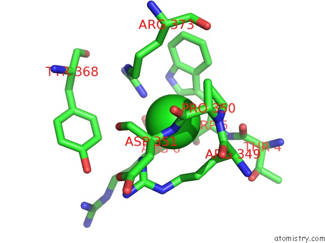
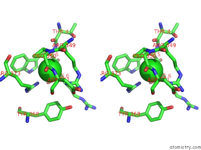
Chlorine binding site 1 out of 4 in 4eac
Go back to
Chlorine binding site 1 out
of 4 in the Crystal Structure of Mannonate Dehydratase From Escherichia Coli Strain K12
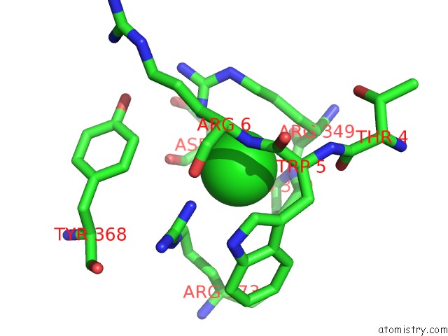
Mono view
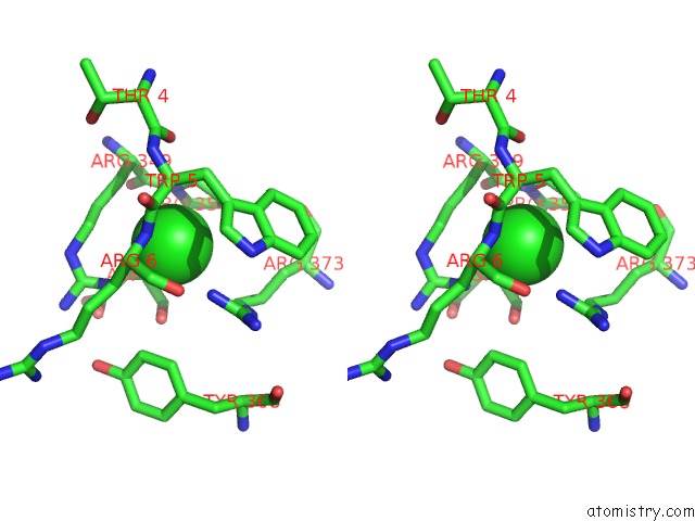
Stereo pair view
Mono view
Stereo pair view
A full contact list of Chlorine with other atoms in the Cl binding
site number 1 of Crystal Structure of Mannonate Dehydratase From Escherichia Coli Strain K12 within 5.0Å range:
|
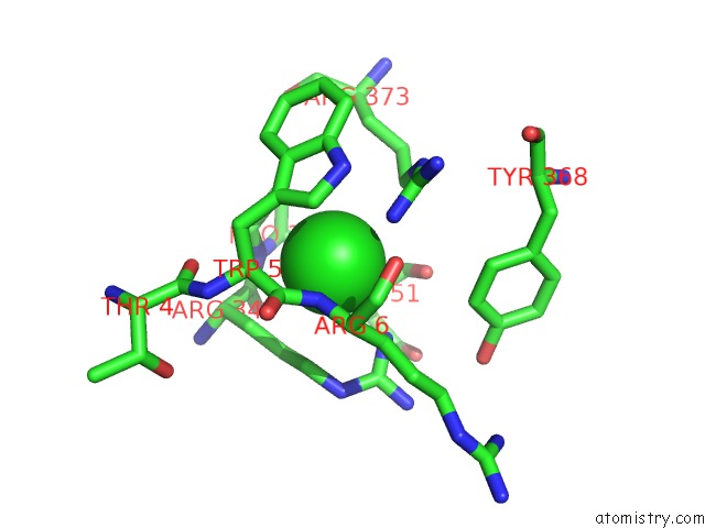
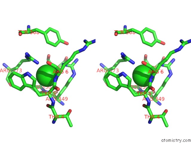
Chlorine binding site 2 out of 4 in 4eac
Go back to
Chlorine binding site 2 out
of 4 in the Crystal Structure of Mannonate Dehydratase From Escherichia Coli Strain K12
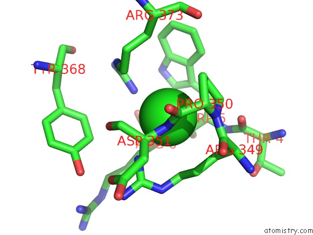
Mono view
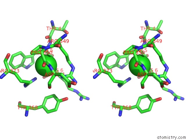
Stereo pair view
Mono view
Stereo pair view
A full contact list of Chlorine with other atoms in the Cl binding
site number 2 of Crystal Structure of Mannonate Dehydratase From Escherichia Coli Strain K12 within 5.0Å range:
|
Chlorine binding site 3 out of 4 in 4eac
Go back to
Chlorine binding site 3 out
of 4 in the Crystal Structure of Mannonate Dehydratase From Escherichia Coli Strain K12
Mono view
Stereo pair view
Mono view
Stereo pair view
A full contact list of Chlorine with other atoms in the Cl binding
site number 3 of Crystal Structure of Mannonate Dehydratase From Escherichia Coli Strain K12 within 5.0Å range:
|
Chlorine binding site 4 out of 4 in 4eac
Go back to
Chlorine binding site 4 out
of 4 in the Crystal Structure of Mannonate Dehydratase From Escherichia Coli Strain K12
Mono view
Stereo pair view
Mono view
Stereo pair view
A full contact list of Chlorine with other atoms in the Cl binding
site number 4 of Crystal Structure of Mannonate Dehydratase From Escherichia Coli Strain K12 within 5.0Å range:
|
Reference:
X.Qiu,
Y.Tao,
Y.Zhu,
Y.Yuan,
Y.Zhang,
H.Liu,
Y.Gao,
M.Teng,
L.Niu.
Structural Insights Into Decreased Enzymatic Activity Induced By An Insert Sequence in Mannonate Dehydratase From Gram Negative Bacterium. J.Struct.Biol. V. 180 327 2012.
ISSN: ISSN 1047-8477
PubMed: 22796868
DOI: 10.1016/J.JSB.2012.06.013
Page generated: Sun Jul 21 12:42:19 2024
ISSN: ISSN 1047-8477
PubMed: 22796868
DOI: 10.1016/J.JSB.2012.06.013
Last articles
Zn in 9JYWZn in 9IR4
Zn in 9IR3
Zn in 9GMX
Zn in 9GMW
Zn in 9JEJ
Zn in 9ERF
Zn in 9ERE
Zn in 9EGV
Zn in 9EGW