Chlorine »
PDB 4egp-4emw »
4ehy »
Chlorine in PDB 4ehy: Crystal Structure of Lpxk From Aquifex Aeolicus in Complex with Adp/MG2+ at 2.2 Angstrom Resolution
Enzymatic activity of Crystal Structure of Lpxk From Aquifex Aeolicus in Complex with Adp/MG2+ at 2.2 Angstrom Resolution
All present enzymatic activity of Crystal Structure of Lpxk From Aquifex Aeolicus in Complex with Adp/MG2+ at 2.2 Angstrom Resolution:
2.7.1.130;
2.7.1.130;
Protein crystallography data
The structure of Crystal Structure of Lpxk From Aquifex Aeolicus in Complex with Adp/MG2+ at 2.2 Angstrom Resolution, PDB code: 4ehy
was solved by
R.P.Emptage,
K.D.Daughtry,
C.W.Pemble Iv,
C.R.H.Raetz,
with X-Ray Crystallography technique. A brief refinement statistics is given in the table below:
| Resolution Low / High (Å) | 28.40 / 2.20 |
| Space group | P 21 21 21 |
| Cell size a, b, c (Å), α, β, γ (°) | 65.110, 75.610, 104.200, 90.00, 90.00, 90.00 |
| R / Rfree (%) | 17.8 / 21.3 |
Other elements in 4ehy:
The structure of Crystal Structure of Lpxk From Aquifex Aeolicus in Complex with Adp/MG2+ at 2.2 Angstrom Resolution also contains other interesting chemical elements:
| Magnesium | (Mg) | 1 atom |
Chlorine Binding Sites:
The binding sites of Chlorine atom in the Crystal Structure of Lpxk From Aquifex Aeolicus in Complex with Adp/MG2+ at 2.2 Angstrom Resolution
(pdb code 4ehy). This binding sites where shown within
5.0 Angstroms radius around Chlorine atom.
In total only one binding site of Chlorine was determined in the Crystal Structure of Lpxk From Aquifex Aeolicus in Complex with Adp/MG2+ at 2.2 Angstrom Resolution, PDB code: 4ehy:
In total only one binding site of Chlorine was determined in the Crystal Structure of Lpxk From Aquifex Aeolicus in Complex with Adp/MG2+ at 2.2 Angstrom Resolution, PDB code: 4ehy:
Chlorine binding site 1 out of 1 in 4ehy
Go back to
Chlorine binding site 1 out
of 1 in the Crystal Structure of Lpxk From Aquifex Aeolicus in Complex with Adp/MG2+ at 2.2 Angstrom Resolution
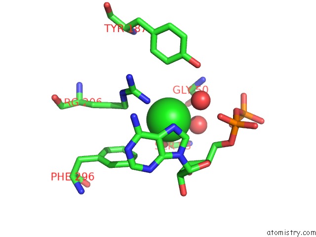
Mono view
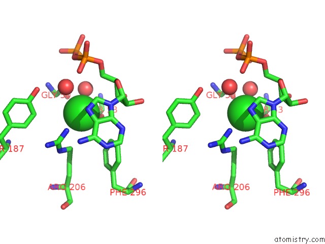
Stereo pair view
Mono view
Stereo pair view
A full contact list of Chlorine with other atoms in the Cl binding
site number 1 of Crystal Structure of Lpxk From Aquifex Aeolicus in Complex with Adp/MG2+ at 2.2 Angstrom Resolution within 5.0Å range:
|
Reference:
R.P.Emptage,
K.D.Daughtry,
C.W.Pemble,
C.R.Raetz.
Crystal Structure of Lpxk, the 4'-Kinase of Lipid A Biosynthesis and Atypical P-Loop Kinase Functioning at the Membrane Interface. Proc.Natl.Acad.Sci.Usa V. 109 12956 2012.
ISSN: ISSN 0027-8424
PubMed: 22826246
DOI: 10.1073/PNAS.1206072109
Page generated: Fri Jul 11 14:50:37 2025
ISSN: ISSN 0027-8424
PubMed: 22826246
DOI: 10.1073/PNAS.1206072109
Last articles
Cl in 8CIGCl in 8CI3
Cl in 8CHM
Cl in 8CHY
Cl in 8CHR
Cl in 8CHQ
Cl in 8CHP
Cl in 8CHL
Cl in 8CHJ
Cl in 8CHN