Chlorine »
PDB 4f1b-4fbx »
4f8h »
Chlorine in PDB 4f8h: X-Ray Structure of the Anesthetic Ketamine Bound to the Glic Pentameric Ligand-Gated Ion Channel
Protein crystallography data
The structure of X-Ray Structure of the Anesthetic Ketamine Bound to the Glic Pentameric Ligand-Gated Ion Channel, PDB code: 4f8h
was solved by
J.J.Pan,
Q.Chen,
D.Willenbring,
X.P.Kong,
A.Cohen,
Y.Xu,
P.Tang,
with X-Ray Crystallography technique. A brief refinement statistics is given in the table below:
| Resolution Low / High (Å) | 24.84 / 2.99 |
| Space group | C 1 2 1 |
| Cell size a, b, c (Å), α, β, γ (°) | 184.069, 132.742, 162.077, 90.00, 103.61, 90.00 |
| R / Rfree (%) | 18.7 / 21.9 |
Chlorine Binding Sites:
The binding sites of Chlorine atom in the X-Ray Structure of the Anesthetic Ketamine Bound to the Glic Pentameric Ligand-Gated Ion Channel
(pdb code 4f8h). This binding sites where shown within
5.0 Angstroms radius around Chlorine atom.
In total 5 binding sites of Chlorine where determined in the X-Ray Structure of the Anesthetic Ketamine Bound to the Glic Pentameric Ligand-Gated Ion Channel, PDB code: 4f8h:
Jump to Chlorine binding site number: 1; 2; 3; 4; 5;
In total 5 binding sites of Chlorine where determined in the X-Ray Structure of the Anesthetic Ketamine Bound to the Glic Pentameric Ligand-Gated Ion Channel, PDB code: 4f8h:
Jump to Chlorine binding site number: 1; 2; 3; 4; 5;
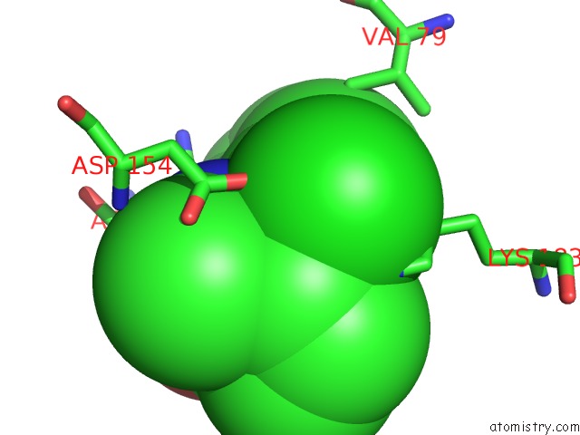
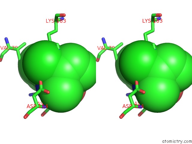
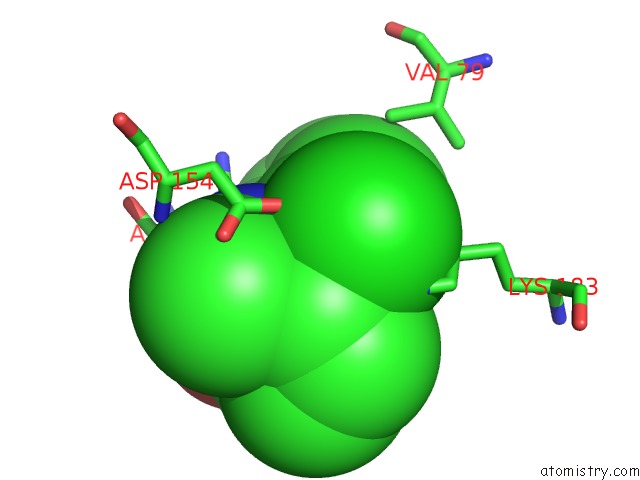
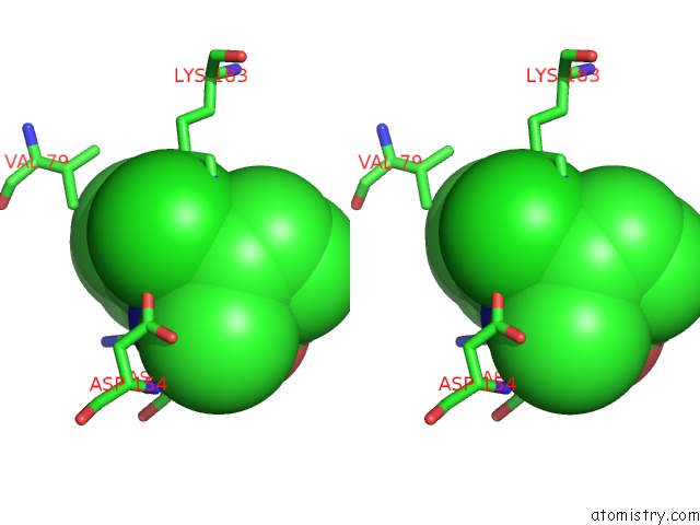
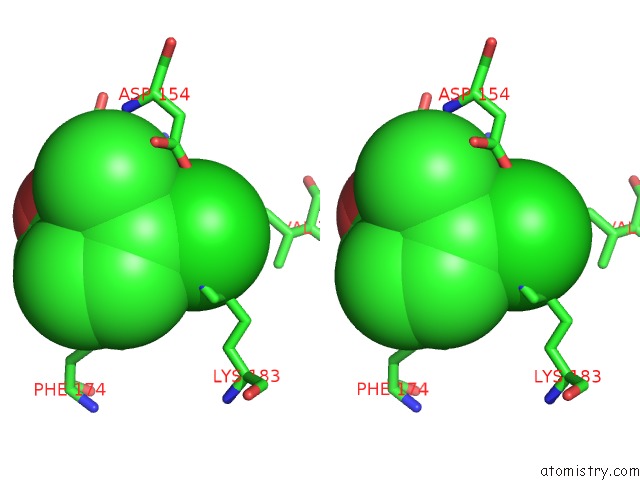
Chlorine binding site 1 out of 5 in 4f8h
Go back to
Chlorine binding site 1 out
of 5 in the X-Ray Structure of the Anesthetic Ketamine Bound to the Glic Pentameric Ligand-Gated Ion Channel
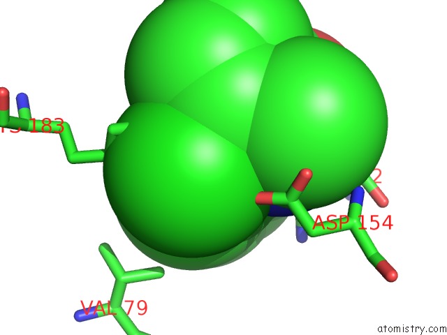
Mono view

Stereo pair view
Mono view
Stereo pair view
A full contact list of Chlorine with other atoms in the Cl binding
site number 1 of X-Ray Structure of the Anesthetic Ketamine Bound to the Glic Pentameric Ligand-Gated Ion Channel within 5.0Å range:
|
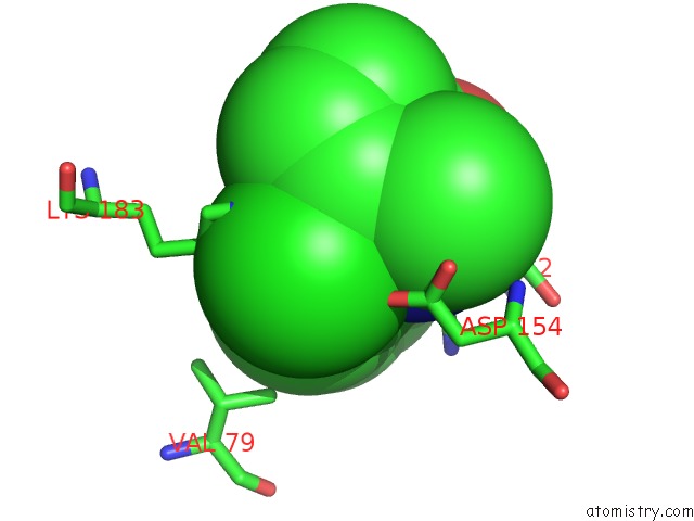
Chlorine binding site 2 out of 5 in 4f8h
Go back to
Chlorine binding site 2 out
of 5 in the X-Ray Structure of the Anesthetic Ketamine Bound to the Glic Pentameric Ligand-Gated Ion Channel
Mono view
Stereo pair view
Mono view
Stereo pair view
A full contact list of Chlorine with other atoms in the Cl binding
site number 2 of X-Ray Structure of the Anesthetic Ketamine Bound to the Glic Pentameric Ligand-Gated Ion Channel within 5.0Å range:
|
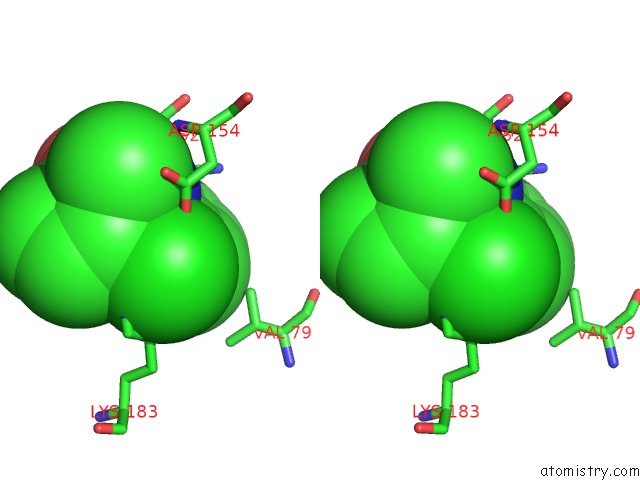
Chlorine binding site 3 out of 5 in 4f8h
Go back to
Chlorine binding site 3 out
of 5 in the X-Ray Structure of the Anesthetic Ketamine Bound to the Glic Pentameric Ligand-Gated Ion Channel
Mono view
Stereo pair view
Mono view
Stereo pair view
A full contact list of Chlorine with other atoms in the Cl binding
site number 3 of X-Ray Structure of the Anesthetic Ketamine Bound to the Glic Pentameric Ligand-Gated Ion Channel within 5.0Å range:
|
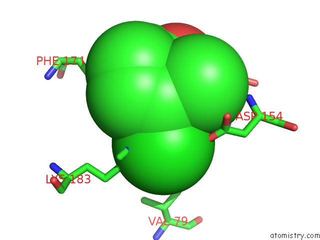
Chlorine binding site 4 out of 5 in 4f8h
Go back to
Chlorine binding site 4 out
of 5 in the X-Ray Structure of the Anesthetic Ketamine Bound to the Glic Pentameric Ligand-Gated Ion Channel
Mono view
Stereo pair view
Mono view
Stereo pair view
A full contact list of Chlorine with other atoms in the Cl binding
site number 4 of X-Ray Structure of the Anesthetic Ketamine Bound to the Glic Pentameric Ligand-Gated Ion Channel within 5.0Å range:
|
Chlorine binding site 5 out of 5 in 4f8h
Go back to
Chlorine binding site 5 out
of 5 in the X-Ray Structure of the Anesthetic Ketamine Bound to the Glic Pentameric Ligand-Gated Ion Channel
Mono view
Stereo pair view
Mono view
Stereo pair view
A full contact list of Chlorine with other atoms in the Cl binding
site number 5 of X-Ray Structure of the Anesthetic Ketamine Bound to the Glic Pentameric Ligand-Gated Ion Channel within 5.0Å range:
|
Reference:
J.Pan,
Q.Chen,
D.Willenbring,
D.Mowrey,
X.P.Kong,
A.Cohen,
C.B.Divito,
Y.Xu,
P.Tang.
Structure of the Pentameric Ligand-Gated Ion Channel Glic Bound with Anesthetic Ketamine. Structure V. 20 1463 2012.
ISSN: ISSN 0969-2126
PubMed: 22958642
DOI: 10.1016/J.STR.2012.08.009
Page generated: Sun Jul 21 13:35:24 2024
ISSN: ISSN 0969-2126
PubMed: 22958642
DOI: 10.1016/J.STR.2012.08.009
Last articles
Zn in 9J0NZn in 9J0O
Zn in 9J0P
Zn in 9FJX
Zn in 9EKB
Zn in 9C0F
Zn in 9CAH
Zn in 9CH0
Zn in 9CH3
Zn in 9CH1