Chlorine »
PDB 4fby-4fm6 »
4fe2 »
Chlorine in PDB 4fe2: X-Ray Structure of Saicar Synthetase (Purc) From Streptococcus Pneumoniae Complexed with Air, Adp, Asp and MG2+
Enzymatic activity of X-Ray Structure of Saicar Synthetase (Purc) From Streptococcus Pneumoniae Complexed with Air, Adp, Asp and MG2+
All present enzymatic activity of X-Ray Structure of Saicar Synthetase (Purc) From Streptococcus Pneumoniae Complexed with Air, Adp, Asp and MG2+:
6.3.2.6;
6.3.2.6;
Protein crystallography data
The structure of X-Ray Structure of Saicar Synthetase (Purc) From Streptococcus Pneumoniae Complexed with Air, Adp, Asp and MG2+, PDB code: 4fe2
was solved by
N.Wolf,
C.Abad-Zapatero,
M.E.Johnson,
L.M.-W.Fung,
with X-Ray Crystallography technique. A brief refinement statistics is given in the table below:
| Resolution Low / High (Å) | 85.99 / 2.29 |
| Space group | P 1 21 1 |
| Cell size a, b, c (Å), α, β, γ (°) | 46.074, 65.021, 86.052, 90.00, 92.09, 90.00 |
| R / Rfree (%) | 19.6 / 28.4 |
Other elements in 4fe2:
The structure of X-Ray Structure of Saicar Synthetase (Purc) From Streptococcus Pneumoniae Complexed with Air, Adp, Asp and MG2+ also contains other interesting chemical elements:
| Magnesium | (Mg) | 2 atoms |
Chlorine Binding Sites:
The binding sites of Chlorine atom in the X-Ray Structure of Saicar Synthetase (Purc) From Streptococcus Pneumoniae Complexed with Air, Adp, Asp and MG2+
(pdb code 4fe2). This binding sites where shown within
5.0 Angstroms radius around Chlorine atom.
In total only one binding site of Chlorine was determined in the X-Ray Structure of Saicar Synthetase (Purc) From Streptococcus Pneumoniae Complexed with Air, Adp, Asp and MG2+, PDB code: 4fe2:
In total only one binding site of Chlorine was determined in the X-Ray Structure of Saicar Synthetase (Purc) From Streptococcus Pneumoniae Complexed with Air, Adp, Asp and MG2+, PDB code: 4fe2:
Chlorine binding site 1 out of 1 in 4fe2
Go back to
Chlorine binding site 1 out
of 1 in the X-Ray Structure of Saicar Synthetase (Purc) From Streptococcus Pneumoniae Complexed with Air, Adp, Asp and MG2+
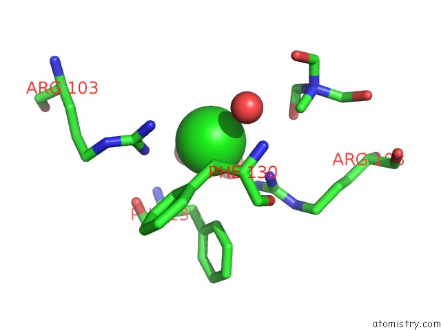
Mono view
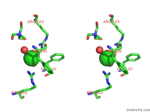
Stereo pair view
Mono view
Stereo pair view
A full contact list of Chlorine with other atoms in the Cl binding
site number 1 of X-Ray Structure of Saicar Synthetase (Purc) From Streptococcus Pneumoniae Complexed with Air, Adp, Asp and MG2+ within 5.0Å range:
|
Reference:
N.M.Wolf,
C.Abad-Zapatero,
M.E.Johnson,
L.W.Fung.
Structures of Saicar Synthetase (Purc) From Streptococcus Pneumoniae with Adp, Mg(2+), Air and Asp. Acta Crystallogr.,Sect.D V. 70 841 2014.
ISSN: ISSN 0907-4449
PubMed: 24598753
DOI: 10.1107/S139900471303366X
Page generated: Sun Jul 21 13:41:25 2024
ISSN: ISSN 0907-4449
PubMed: 24598753
DOI: 10.1107/S139900471303366X
Last articles
Zn in 9J0NZn in 9J0O
Zn in 9J0P
Zn in 9FJX
Zn in 9EKB
Zn in 9C0F
Zn in 9CAH
Zn in 9CH0
Zn in 9CH3
Zn in 9CH1