Chlorine »
PDB 4fv3-4g7y »
4fzl »
Chlorine in PDB 4fzl: High Resolution Structure of Truncated Bacteriocin Syringacin M From Pseudomonas Syringae Pv. Tomato DC3000
Protein crystallography data
The structure of High Resolution Structure of Truncated Bacteriocin Syringacin M From Pseudomonas Syringae Pv. Tomato DC3000, PDB code: 4fzl
was solved by
A.W.Roszak,
R.Grinter,
J.R.Cogdell,
D.Walker,
with X-Ray Crystallography technique. A brief refinement statistics is given in the table below:
| Resolution Low / High (Å) | 50.92 / 1.46 |
| Space group | P 1 21 1 |
| Cell size a, b, c (Å), α, β, γ (°) | 55.810, 96.090, 64.270, 90.00, 114.17, 90.00 |
| R / Rfree (%) | 15.2 / 20.1 |
Other elements in 4fzl:
The structure of High Resolution Structure of Truncated Bacteriocin Syringacin M From Pseudomonas Syringae Pv. Tomato DC3000 also contains other interesting chemical elements:
| Magnesium | (Mg) | 1 atom |
| Calcium | (Ca) | 2 atoms |
Chlorine Binding Sites:
The binding sites of Chlorine atom in the High Resolution Structure of Truncated Bacteriocin Syringacin M From Pseudomonas Syringae Pv. Tomato DC3000
(pdb code 4fzl). This binding sites where shown within
5.0 Angstroms radius around Chlorine atom.
In total 2 binding sites of Chlorine where determined in the High Resolution Structure of Truncated Bacteriocin Syringacin M From Pseudomonas Syringae Pv. Tomato DC3000, PDB code: 4fzl:
Jump to Chlorine binding site number: 1; 2;
In total 2 binding sites of Chlorine where determined in the High Resolution Structure of Truncated Bacteriocin Syringacin M From Pseudomonas Syringae Pv. Tomato DC3000, PDB code: 4fzl:
Jump to Chlorine binding site number: 1; 2;
Chlorine binding site 1 out of 2 in 4fzl
Go back to
Chlorine binding site 1 out
of 2 in the High Resolution Structure of Truncated Bacteriocin Syringacin M From Pseudomonas Syringae Pv. Tomato DC3000
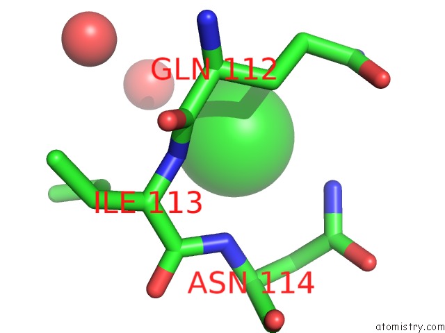
Mono view
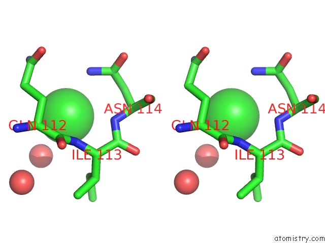
Stereo pair view
Mono view
Stereo pair view
A full contact list of Chlorine with other atoms in the Cl binding
site number 1 of High Resolution Structure of Truncated Bacteriocin Syringacin M From Pseudomonas Syringae Pv. Tomato DC3000 within 5.0Å range:
|
Chlorine binding site 2 out of 2 in 4fzl
Go back to
Chlorine binding site 2 out
of 2 in the High Resolution Structure of Truncated Bacteriocin Syringacin M From Pseudomonas Syringae Pv. Tomato DC3000
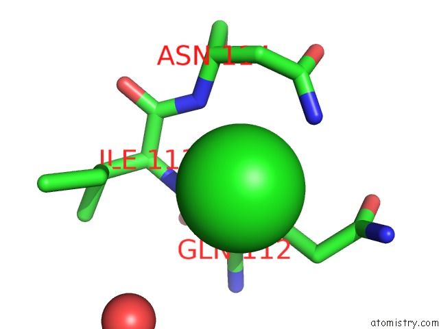
Mono view
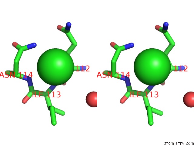
Stereo pair view
Mono view
Stereo pair view
A full contact list of Chlorine with other atoms in the Cl binding
site number 2 of High Resolution Structure of Truncated Bacteriocin Syringacin M From Pseudomonas Syringae Pv. Tomato DC3000 within 5.0Å range:
|
Reference:
R.Grinter,
A.W.Roszak,
R.J.Cogdell,
J.J.Milner,
D.Walker.
The Crystal Structure of the Lipid II-Degrading Bacteriocin Syringacin M Suggests Unexpected Evolutionary Relationships Between Colicin M-Like Bacteriocins. J.Biol.Chem. V. 287 38876 2012.
ISSN: ISSN 0021-9258
PubMed: 22995910
DOI: 10.1074/JBC.M112.400150
Page generated: Sun Jul 21 14:11:26 2024
ISSN: ISSN 0021-9258
PubMed: 22995910
DOI: 10.1074/JBC.M112.400150
Last articles
Zn in 9J0NZn in 9J0O
Zn in 9J0P
Zn in 9FJX
Zn in 9EKB
Zn in 9C0F
Zn in 9CAH
Zn in 9CH0
Zn in 9CH3
Zn in 9CH1