Chlorine »
PDB 4gta-4h1t »
4gze »
Chlorine in PDB 4gze: Crystal Structure of 6-Phospho-Beta-Glucosidase From Lactobacillus Plantarum (Apo Form)
Enzymatic activity of Crystal Structure of 6-Phospho-Beta-Glucosidase From Lactobacillus Plantarum (Apo Form)
All present enzymatic activity of Crystal Structure of 6-Phospho-Beta-Glucosidase From Lactobacillus Plantarum (Apo Form):
3.2.1.86;
3.2.1.86;
Protein crystallography data
The structure of Crystal Structure of 6-Phospho-Beta-Glucosidase From Lactobacillus Plantarum (Apo Form), PDB code: 4gze
was solved by
K.Michalska,
C.Hatzos-Skintges,
J.Bearden,
A.Joachimiak,
Midwest Centerfor Structural Genomics (Mcsg),
with X-Ray Crystallography technique. A brief refinement statistics is given in the table below:
| Resolution Low / High (Å) | 30.00 / 2.31 |
| Space group | P 31 |
| Cell size a, b, c (Å), α, β, γ (°) | 96.086, 96.086, 289.140, 90.00, 90.00, 120.00 |
| R / Rfree (%) | 17.4 / 21.3 |
Chlorine Binding Sites:
The binding sites of Chlorine atom in the Crystal Structure of 6-Phospho-Beta-Glucosidase From Lactobacillus Plantarum (Apo Form)
(pdb code 4gze). This binding sites where shown within
5.0 Angstroms radius around Chlorine atom.
In total 6 binding sites of Chlorine where determined in the Crystal Structure of 6-Phospho-Beta-Glucosidase From Lactobacillus Plantarum (Apo Form), PDB code: 4gze:
Jump to Chlorine binding site number: 1; 2; 3; 4; 5; 6;
In total 6 binding sites of Chlorine where determined in the Crystal Structure of 6-Phospho-Beta-Glucosidase From Lactobacillus Plantarum (Apo Form), PDB code: 4gze:
Jump to Chlorine binding site number: 1; 2; 3; 4; 5; 6;
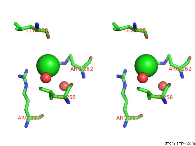
Chlorine binding site 1 out of 6 in 4gze
Go back to
Chlorine binding site 1 out
of 6 in the Crystal Structure of 6-Phospho-Beta-Glucosidase From Lactobacillus Plantarum (Apo Form)

Mono view

Stereo pair view
Mono view
Stereo pair view
A full contact list of Chlorine with other atoms in the Cl binding
site number 1 of Crystal Structure of 6-Phospho-Beta-Glucosidase From Lactobacillus Plantarum (Apo Form) within 5.0Å range:
|
Chlorine binding site 2 out of 6 in 4gze
Go back to
Chlorine binding site 2 out
of 6 in the Crystal Structure of 6-Phospho-Beta-Glucosidase From Lactobacillus Plantarum (Apo Form)

Mono view

Stereo pair view
Mono view
Stereo pair view
A full contact list of Chlorine with other atoms in the Cl binding
site number 2 of Crystal Structure of 6-Phospho-Beta-Glucosidase From Lactobacillus Plantarum (Apo Form) within 5.0Å range:
|
Chlorine binding site 3 out of 6 in 4gze
Go back to
Chlorine binding site 3 out
of 6 in the Crystal Structure of 6-Phospho-Beta-Glucosidase From Lactobacillus Plantarum (Apo Form)

Mono view

Stereo pair view
Mono view
Stereo pair view
A full contact list of Chlorine with other atoms in the Cl binding
site number 3 of Crystal Structure of 6-Phospho-Beta-Glucosidase From Lactobacillus Plantarum (Apo Form) within 5.0Å range:
|
Chlorine binding site 4 out of 6 in 4gze
Go back to
Chlorine binding site 4 out
of 6 in the Crystal Structure of 6-Phospho-Beta-Glucosidase From Lactobacillus Plantarum (Apo Form)

Mono view
Stereo pair view
Mono view
Stereo pair view
A full contact list of Chlorine with other atoms in the Cl binding
site number 4 of Crystal Structure of 6-Phospho-Beta-Glucosidase From Lactobacillus Plantarum (Apo Form) within 5.0Å range:
|
Chlorine binding site 5 out of 6 in 4gze
Go back to
Chlorine binding site 5 out
of 6 in the Crystal Structure of 6-Phospho-Beta-Glucosidase From Lactobacillus Plantarum (Apo Form)

Mono view

Stereo pair view
Mono view
Stereo pair view
A full contact list of Chlorine with other atoms in the Cl binding
site number 5 of Crystal Structure of 6-Phospho-Beta-Glucosidase From Lactobacillus Plantarum (Apo Form) within 5.0Å range:
|
Chlorine binding site 6 out of 6 in 4gze
Go back to
Chlorine binding site 6 out
of 6 in the Crystal Structure of 6-Phospho-Beta-Glucosidase From Lactobacillus Plantarum (Apo Form)

Mono view

Stereo pair view
Mono view
Stereo pair view
A full contact list of Chlorine with other atoms in the Cl binding
site number 6 of Crystal Structure of 6-Phospho-Beta-Glucosidase From Lactobacillus Plantarum (Apo Form) within 5.0Å range:
|
Reference:
K.Michalska,
K.Tan,
H.Li,
C.Hatzos-Skintges,
J.Bearden,
G.Babnigg,
A.Joachimiak.
GH1-Family 6-P-Beta-Glucosidases From Human Microbiome Lactic Acid Bacteria. Acta Crystallogr.,Sect.D V. 69 451 2013.
ISSN: ISSN 0907-4449
PubMed: 23519420
DOI: 10.1107/S0907444912049608
Page generated: Sun Jul 21 15:11:54 2024
ISSN: ISSN 0907-4449
PubMed: 23519420
DOI: 10.1107/S0907444912049608
Last articles
Zn in 9J0NZn in 9J0O
Zn in 9J0P
Zn in 9FJX
Zn in 9EKB
Zn in 9C0F
Zn in 9CAH
Zn in 9CH0
Zn in 9CH3
Zn in 9CH1