Chlorine »
PDB 4h1x-4h8x »
4h3z »
Chlorine in PDB 4h3z: Crystal Structure of A Symmetric Dimer of A Trna (Guanine-(N(1)-)- Methyltransferase From Burkholderia Phymatum Bound to S-Adenosyl Homocystein in Both Half-Sites
Enzymatic activity of Crystal Structure of A Symmetric Dimer of A Trna (Guanine-(N(1)-)- Methyltransferase From Burkholderia Phymatum Bound to S-Adenosyl Homocystein in Both Half-Sites
All present enzymatic activity of Crystal Structure of A Symmetric Dimer of A Trna (Guanine-(N(1)-)- Methyltransferase From Burkholderia Phymatum Bound to S-Adenosyl Homocystein in Both Half-Sites:
2.1.1.228;
2.1.1.228;
Protein crystallography data
The structure of Crystal Structure of A Symmetric Dimer of A Trna (Guanine-(N(1)-)- Methyltransferase From Burkholderia Phymatum Bound to S-Adenosyl Homocystein in Both Half-Sites, PDB code: 4h3z
was solved by
Seattle Structural Genomics Center For Infectious Disease (Ssgcid),
with X-Ray Crystallography technique. A brief refinement statistics is given in the table below:
| Resolution Low / High (Å) | 42.20 / 2.15 |
| Space group | C 2 2 21 |
| Cell size a, b, c (Å), α, β, γ (°) | 58.850, 188.090, 127.680, 90.00, 90.00, 90.00 |
| R / Rfree (%) | 18.6 / 22.8 |
Chlorine Binding Sites:
The binding sites of Chlorine atom in the Crystal Structure of A Symmetric Dimer of A Trna (Guanine-(N(1)-)- Methyltransferase From Burkholderia Phymatum Bound to S-Adenosyl Homocystein in Both Half-Sites
(pdb code 4h3z). This binding sites where shown within
5.0 Angstroms radius around Chlorine atom.
In total 2 binding sites of Chlorine where determined in the Crystal Structure of A Symmetric Dimer of A Trna (Guanine-(N(1)-)- Methyltransferase From Burkholderia Phymatum Bound to S-Adenosyl Homocystein in Both Half-Sites, PDB code: 4h3z:
Jump to Chlorine binding site number: 1; 2;
In total 2 binding sites of Chlorine where determined in the Crystal Structure of A Symmetric Dimer of A Trna (Guanine-(N(1)-)- Methyltransferase From Burkholderia Phymatum Bound to S-Adenosyl Homocystein in Both Half-Sites, PDB code: 4h3z:
Jump to Chlorine binding site number: 1; 2;
Chlorine binding site 1 out of 2 in 4h3z
Go back to
Chlorine binding site 1 out
of 2 in the Crystal Structure of A Symmetric Dimer of A Trna (Guanine-(N(1)-)- Methyltransferase From Burkholderia Phymatum Bound to S-Adenosyl Homocystein in Both Half-Sites
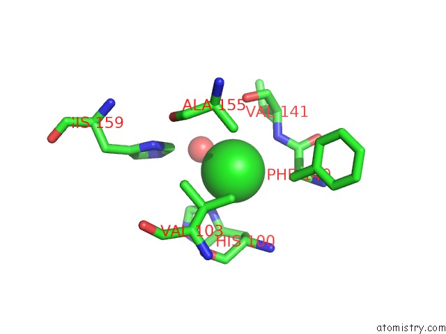
Mono view
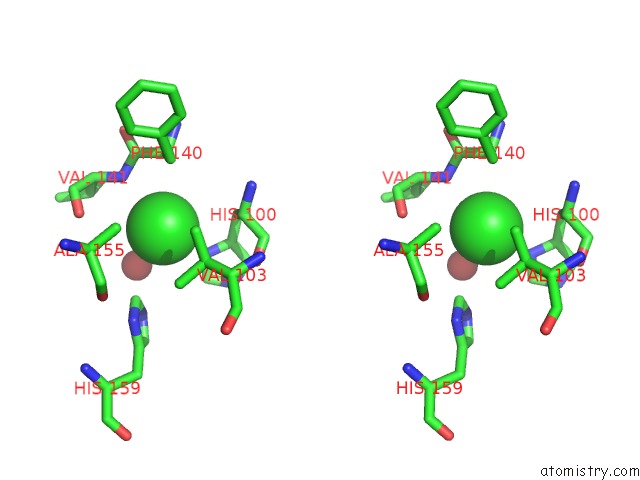
Stereo pair view
Mono view
Stereo pair view
A full contact list of Chlorine with other atoms in the Cl binding
site number 1 of Crystal Structure of A Symmetric Dimer of A Trna (Guanine-(N(1)-)- Methyltransferase From Burkholderia Phymatum Bound to S-Adenosyl Homocystein in Both Half-Sites within 5.0Å range:
|
Chlorine binding site 2 out of 2 in 4h3z
Go back to
Chlorine binding site 2 out
of 2 in the Crystal Structure of A Symmetric Dimer of A Trna (Guanine-(N(1)-)- Methyltransferase From Burkholderia Phymatum Bound to S-Adenosyl Homocystein in Both Half-Sites
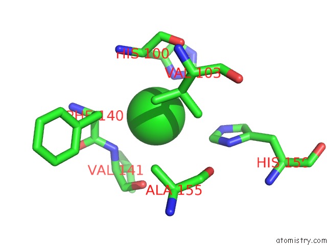
Mono view
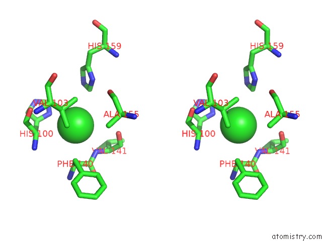
Stereo pair view
Mono view
Stereo pair view
A full contact list of Chlorine with other atoms in the Cl binding
site number 2 of Crystal Structure of A Symmetric Dimer of A Trna (Guanine-(N(1)-)- Methyltransferase From Burkholderia Phymatum Bound to S-Adenosyl Homocystein in Both Half-Sites within 5.0Å range:
|
Reference:
L.Baugh,
L.A.Gallagher,
R.Patrapuvich,
M.C.Clifton,
A.S.Gardberg,
T.E.Edwards,
B.Armour,
D.W.Begley,
S.H.Dieterich,
D.M.Dranow,
J.Abendroth,
J.W.Fairman,
D.Fox,
B.L.Staker,
I.Phan,
A.Gillespie,
R.Choi,
S.Nakazawa-Hewitt,
M.T.Nguyen,
A.Napuli,
L.Barrett,
G.W.Buchko,
R.Stacy,
P.J.Myler,
L.J.Stewart,
C.Manoil,
W.C.Van Voorhis.
Combining Functional and Structural Genomics to Sample the Essential Burkholderia Structome. Plos One V. 8 53851 2013.
ISSN: ESSN 1932-6203
PubMed: 23382856
DOI: 10.1371/JOURNAL.PONE.0053851
Page generated: Sun Jul 21 15:26:19 2024
ISSN: ESSN 1932-6203
PubMed: 23382856
DOI: 10.1371/JOURNAL.PONE.0053851
Last articles
Ca in 5NYYCa in 5NXL
Ca in 5NXU
Ca in 5NXR
Ca in 5NXB
Ca in 5NXN
Ca in 5NRK
Ca in 5NUR
Ca in 5NSF
Ca in 5NRM