Chlorine »
PDB 4hn0-4hw2 »
4huf »
Chlorine in PDB 4huf: Structure of 5-Chlorouracil Modified A:U Base Pair
Enzymatic activity of Structure of 5-Chlorouracil Modified A:U Base Pair
All present enzymatic activity of Structure of 5-Chlorouracil Modified A:U Base Pair:
3.1.26.4;
3.1.26.4;
Protein crystallography data
The structure of Structure of 5-Chlorouracil Modified A:U Base Pair, PDB code: 4huf
was solved by
A.Patra,
M.Egli,
with X-Ray Crystallography technique. A brief refinement statistics is given in the table below:
| Resolution Low / High (Å) | 35.90 / 1.69 |
| Space group | P 21 21 21 |
| Cell size a, b, c (Å), α, β, γ (°) | 64.193, 64.748, 116.470, 90.00, 90.00, 90.00 |
| R / Rfree (%) | 19.2 / 22.8 |
Other elements in 4huf:
The structure of Structure of 5-Chlorouracil Modified A:U Base Pair also contains other interesting chemical elements:
| Magnesium | (Mg) | 2 atoms |
Chlorine Binding Sites:
The binding sites of Chlorine atom in the Structure of 5-Chlorouracil Modified A:U Base Pair
(pdb code 4huf). This binding sites where shown within
5.0 Angstroms radius around Chlorine atom.
In total 4 binding sites of Chlorine where determined in the Structure of 5-Chlorouracil Modified A:U Base Pair, PDB code: 4huf:
Jump to Chlorine binding site number: 1; 2; 3; 4;
In total 4 binding sites of Chlorine where determined in the Structure of 5-Chlorouracil Modified A:U Base Pair, PDB code: 4huf:
Jump to Chlorine binding site number: 1; 2; 3; 4;
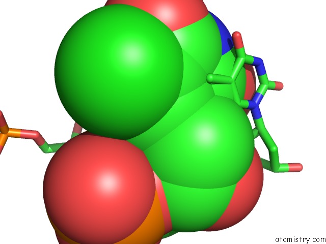
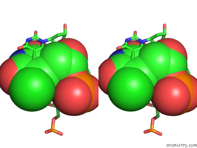
Chlorine binding site 1 out of 4 in 4huf
Go back to
Chlorine binding site 1 out
of 4 in the Structure of 5-Chlorouracil Modified A:U Base Pair
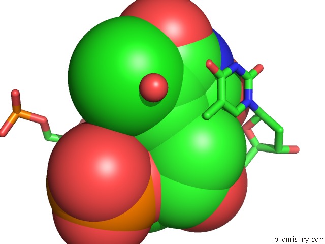
Mono view
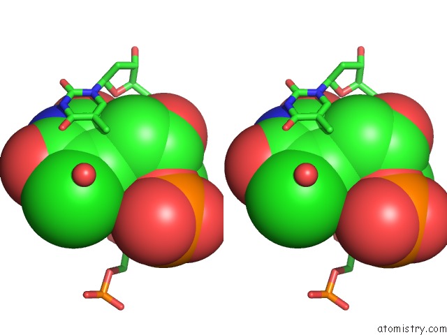
Stereo pair view
Mono view
Stereo pair view
A full contact list of Chlorine with other atoms in the Cl binding
site number 1 of Structure of 5-Chlorouracil Modified A:U Base Pair within 5.0Å range:
|
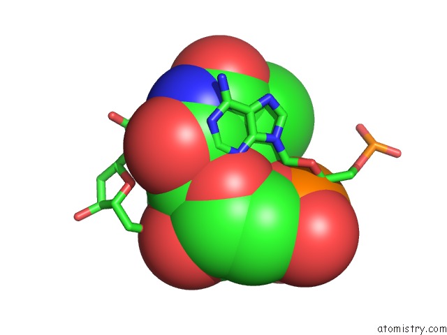
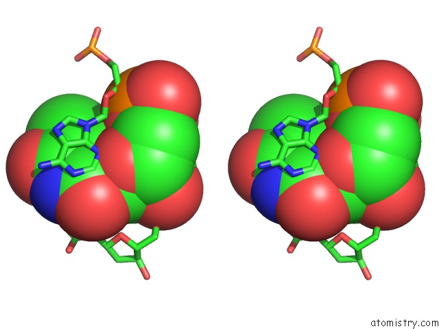
Chlorine binding site 2 out of 4 in 4huf
Go back to
Chlorine binding site 2 out
of 4 in the Structure of 5-Chlorouracil Modified A:U Base Pair
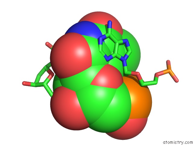
Mono view
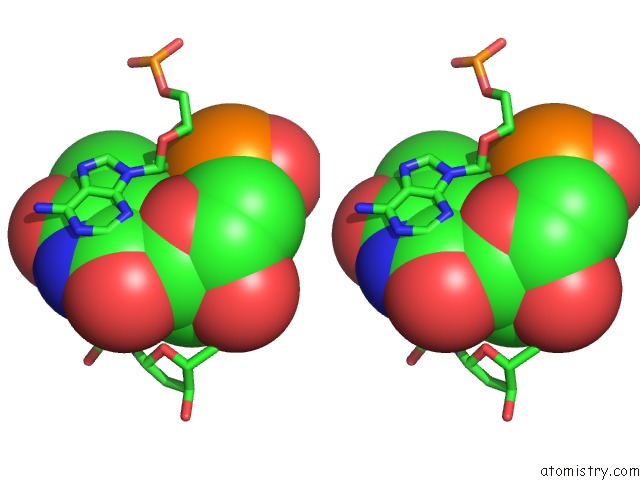
Stereo pair view
Mono view
Stereo pair view
A full contact list of Chlorine with other atoms in the Cl binding
site number 2 of Structure of 5-Chlorouracil Modified A:U Base Pair within 5.0Å range:
|
Chlorine binding site 3 out of 4 in 4huf
Go back to
Chlorine binding site 3 out
of 4 in the Structure of 5-Chlorouracil Modified A:U Base Pair
Mono view
Stereo pair view
Mono view
Stereo pair view
A full contact list of Chlorine with other atoms in the Cl binding
site number 3 of Structure of 5-Chlorouracil Modified A:U Base Pair within 5.0Å range:
|
Chlorine binding site 4 out of 4 in 4huf
Go back to
Chlorine binding site 4 out
of 4 in the Structure of 5-Chlorouracil Modified A:U Base Pair
Mono view
Stereo pair view
Mono view
Stereo pair view
A full contact list of Chlorine with other atoms in the Cl binding
site number 4 of Structure of 5-Chlorouracil Modified A:U Base Pair within 5.0Å range:
|
Reference:
A.Patra,
J.Harp,
P.S.Pallan,
L.Zhao,
M.Abramov,
P.Herdewijn,
M.Egli.
Structure, Stability and Function of 5-Chlorouracil Modified A:U and G:U Base Pairs. Nucleic Acids Res. V. 41 2689 2013.
ISSN: ISSN 0305-1048
PubMed: 23275540
DOI: 10.1093/NAR/GKS1316
Page generated: Sun Jul 21 16:07:01 2024
ISSN: ISSN 0305-1048
PubMed: 23275540
DOI: 10.1093/NAR/GKS1316
Last articles
Zn in 9J0NZn in 9J0O
Zn in 9J0P
Zn in 9FJX
Zn in 9EKB
Zn in 9C0F
Zn in 9CAH
Zn in 9CH0
Zn in 9CH3
Zn in 9CH1