Chlorine »
PDB 4i22-4ibe »
4i50 »
Chlorine in PDB 4i50: Crystal Structure of Tubulin-Stathmin-Ttl-Epothilone A Complex
Protein crystallography data
The structure of Crystal Structure of Tubulin-Stathmin-Ttl-Epothilone A Complex, PDB code: 4i50
was solved by
A.E.Prota,
K.Bargsten,
D.Zurwerra,
J.J.Field,
J.F.Diaz,
K.H.Altmann,
M.O.Steinmetz,
with X-Ray Crystallography technique. A brief refinement statistics is given in the table below:
| Resolution Low / High (Å) | 77.57 / 2.30 |
| Space group | P 21 21 21 |
| Cell size a, b, c (Å), α, β, γ (°) | 103.620, 155.130, 180.410, 90.00, 90.00, 90.00 |
| R / Rfree (%) | 19.1 / 24.6 |
Other elements in 4i50:
The structure of Crystal Structure of Tubulin-Stathmin-Ttl-Epothilone A Complex also contains other interesting chemical elements:
| Magnesium | (Mg) | 6 atoms |
| Calcium | (Ca) | 6 atoms |
Chlorine Binding Sites:
The binding sites of Chlorine atom in the Crystal Structure of Tubulin-Stathmin-Ttl-Epothilone A Complex
(pdb code 4i50). This binding sites where shown within
5.0 Angstroms radius around Chlorine atom.
In total only one binding site of Chlorine was determined in the Crystal Structure of Tubulin-Stathmin-Ttl-Epothilone A Complex, PDB code: 4i50:
In total only one binding site of Chlorine was determined in the Crystal Structure of Tubulin-Stathmin-Ttl-Epothilone A Complex, PDB code: 4i50:
Chlorine binding site 1 out of 1 in 4i50
Go back to
Chlorine binding site 1 out
of 1 in the Crystal Structure of Tubulin-Stathmin-Ttl-Epothilone A Complex
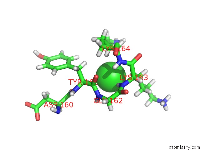
Mono view
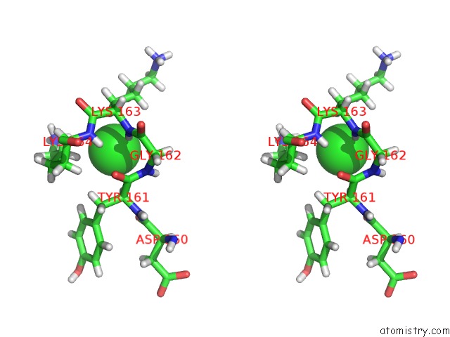
Stereo pair view
Mono view
Stereo pair view
A full contact list of Chlorine with other atoms in the Cl binding
site number 1 of Crystal Structure of Tubulin-Stathmin-Ttl-Epothilone A Complex within 5.0Å range:
|
Reference:
A.E.Prota,
K.Bargsten,
D.Zurwerra,
J.J.Field,
J.F.Diaz,
K.H.Altmann,
M.O.Steinmetz.
Molecular Mechanism of Action of Microtubule-Stabilizing Anticancer Agents. Science V. 339 587 2013.
ISSN: ISSN 0036-8075
PubMed: 23287720
DOI: 10.1126/SCIENCE.1230582
Page generated: Sun Jul 21 16:22:49 2024
ISSN: ISSN 0036-8075
PubMed: 23287720
DOI: 10.1126/SCIENCE.1230582
Last articles
Zn in 9MJ5Zn in 9HNW
Zn in 9G0L
Zn in 9FNE
Zn in 9DZN
Zn in 9E0I
Zn in 9D32
Zn in 9DAK
Zn in 8ZXC
Zn in 8ZUF