Chlorine »
PDB 4i22-4ibe »
4i90 »
Chlorine in PDB 4i90: Structure of the N254Y/H258Y Mutant of the Phosphatidylinositol- Specific Phospholipase C From S. Aureus Bound to Choline
Enzymatic activity of Structure of the N254Y/H258Y Mutant of the Phosphatidylinositol- Specific Phospholipase C From S. Aureus Bound to Choline
All present enzymatic activity of Structure of the N254Y/H258Y Mutant of the Phosphatidylinositol- Specific Phospholipase C From S. Aureus Bound to Choline:
4.6.1.13;
4.6.1.13;
Protein crystallography data
The structure of Structure of the N254Y/H258Y Mutant of the Phosphatidylinositol- Specific Phospholipase C From S. Aureus Bound to Choline, PDB code: 4i90
was solved by
R.I.Goldstein,
J.Cheng,
B.Stec,
A.Gershenson,
M.F.Roberts,
with X-Ray Crystallography technique. A brief refinement statistics is given in the table below:
| Resolution Low / High (Å) | 50.00 / 1.65 |
| Space group | P 21 21 21 |
| Cell size a, b, c (Å), α, β, γ (°) | 85.983, 57.583, 61.694, 90.00, 90.00, 90.00 |
| R / Rfree (%) | 18.5 / 23.2 |
Chlorine Binding Sites:
The binding sites of Chlorine atom in the Structure of the N254Y/H258Y Mutant of the Phosphatidylinositol- Specific Phospholipase C From S. Aureus Bound to Choline
(pdb code 4i90). This binding sites where shown within
5.0 Angstroms radius around Chlorine atom.
In total only one binding site of Chlorine was determined in the Structure of the N254Y/H258Y Mutant of the Phosphatidylinositol- Specific Phospholipase C From S. Aureus Bound to Choline, PDB code: 4i90:
In total only one binding site of Chlorine was determined in the Structure of the N254Y/H258Y Mutant of the Phosphatidylinositol- Specific Phospholipase C From S. Aureus Bound to Choline, PDB code: 4i90:
Chlorine binding site 1 out of 1 in 4i90
Go back to
Chlorine binding site 1 out
of 1 in the Structure of the N254Y/H258Y Mutant of the Phosphatidylinositol- Specific Phospholipase C From S. Aureus Bound to Choline
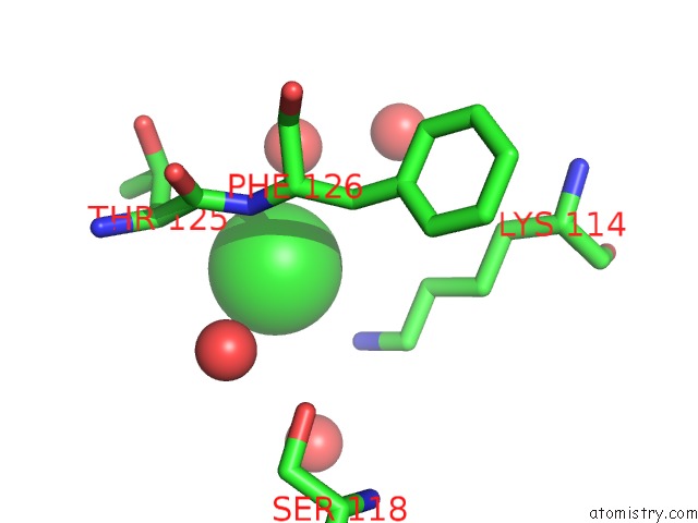
Mono view
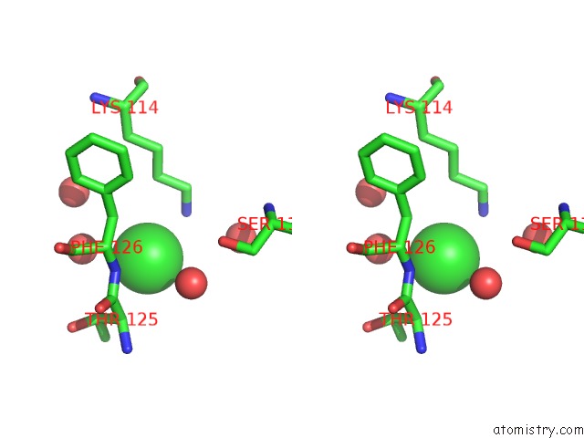
Stereo pair view
Mono view
Stereo pair view
A full contact list of Chlorine with other atoms in the Cl binding
site number 1 of Structure of the N254Y/H258Y Mutant of the Phosphatidylinositol- Specific Phospholipase C From S. Aureus Bound to Choline within 5.0Å range:
|
Reference:
J.Cheng,
R.Goldstein,
A.Gershenson,
B.Stec,
M.F.Roberts.
The Cation-Pi Box Is A Specific Phosphatidylcholine Membrane Targeting Motif. J.Biol.Chem. V. 288 14863 2013.
ISSN: ISSN 0021-9258
PubMed: 23576432
DOI: 10.1074/JBC.M113.466532
Page generated: Sun Jul 21 16:27:36 2024
ISSN: ISSN 0021-9258
PubMed: 23576432
DOI: 10.1074/JBC.M113.466532
Last articles
Zn in 9J0NZn in 9J0O
Zn in 9J0P
Zn in 9FJX
Zn in 9EKB
Zn in 9C0F
Zn in 9CAH
Zn in 9CH0
Zn in 9CH3
Zn in 9CH1