Chlorine »
PDB 4io4-4ixu »
4iwv »
Chlorine in PDB 4iwv: Crystals Structure of Human Glucokinase in Complex with Small Molecule Activator
Enzymatic activity of Crystals Structure of Human Glucokinase in Complex with Small Molecule Activator
All present enzymatic activity of Crystals Structure of Human Glucokinase in Complex with Small Molecule Activator:
2.7.1.2;
2.7.1.2;
Protein crystallography data
The structure of Crystals Structure of Human Glucokinase in Complex with Small Molecule Activator, PDB code: 4iwv
was solved by
D.J.Ogg,
D.Hargreaves,
S.Gerhardt,
L.Flavell,
M.Mcalister,
with X-Ray Crystallography technique. A brief refinement statistics is given in the table below:
| Resolution Low / High (Å) | 37.46 / 2.10 |
| Space group | P 41 |
| Cell size a, b, c (Å), α, β, γ (°) | 77.938, 77.938, 85.350, 90.00, 90.00, 90.00 |
| R / Rfree (%) | 19.9 / 25 |
Other elements in 4iwv:
The structure of Crystals Structure of Human Glucokinase in Complex with Small Molecule Activator also contains other interesting chemical elements:
| Sodium | (Na) | 1 atom |
Chlorine Binding Sites:
The binding sites of Chlorine atom in the Crystals Structure of Human Glucokinase in Complex with Small Molecule Activator
(pdb code 4iwv). This binding sites where shown within
5.0 Angstroms radius around Chlorine atom.
In total 4 binding sites of Chlorine where determined in the Crystals Structure of Human Glucokinase in Complex with Small Molecule Activator, PDB code: 4iwv:
Jump to Chlorine binding site number: 1; 2; 3; 4;
In total 4 binding sites of Chlorine where determined in the Crystals Structure of Human Glucokinase in Complex with Small Molecule Activator, PDB code: 4iwv:
Jump to Chlorine binding site number: 1; 2; 3; 4;
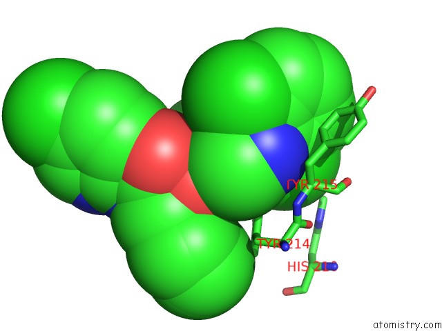
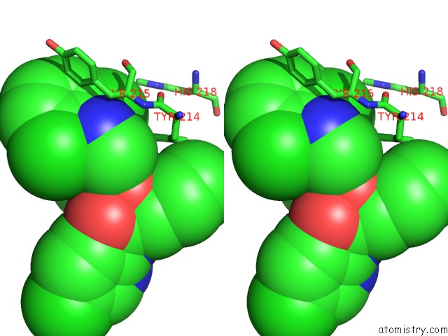
Chlorine binding site 1 out of 4 in 4iwv
Go back to
Chlorine binding site 1 out
of 4 in the Crystals Structure of Human Glucokinase in Complex with Small Molecule Activator

Mono view
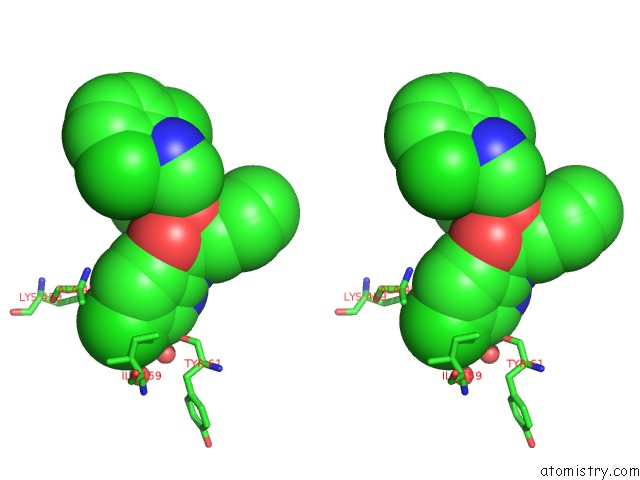
Stereo pair view
Mono view
Stereo pair view
A full contact list of Chlorine with other atoms in the Cl binding
site number 1 of Crystals Structure of Human Glucokinase in Complex with Small Molecule Activator within 5.0Å range:
|
Chlorine binding site 2 out of 4 in 4iwv
Go back to
Chlorine binding site 2 out
of 4 in the Crystals Structure of Human Glucokinase in Complex with Small Molecule Activator
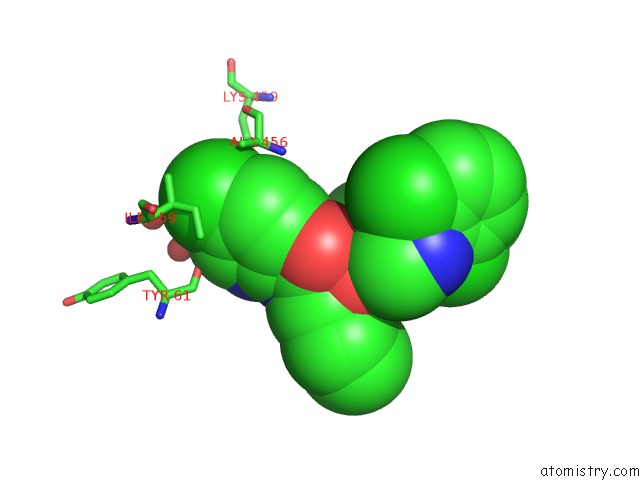
Mono view

Stereo pair view
Mono view
Stereo pair view
A full contact list of Chlorine with other atoms in the Cl binding
site number 2 of Crystals Structure of Human Glucokinase in Complex with Small Molecule Activator within 5.0Å range:
|
Chlorine binding site 3 out of 4 in 4iwv
Go back to
Chlorine binding site 3 out
of 4 in the Crystals Structure of Human Glucokinase in Complex with Small Molecule Activator
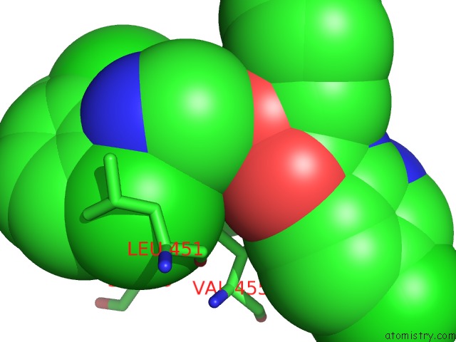
Mono view
Stereo pair view
Mono view
Stereo pair view
A full contact list of Chlorine with other atoms in the Cl binding
site number 3 of Crystals Structure of Human Glucokinase in Complex with Small Molecule Activator within 5.0Å range:
|
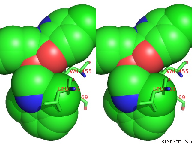
Chlorine binding site 4 out of 4 in 4iwv
Go back to
Chlorine binding site 4 out
of 4 in the Crystals Structure of Human Glucokinase in Complex with Small Molecule Activator
Mono view
Stereo pair view
Mono view
Stereo pair view
A full contact list of Chlorine with other atoms in the Cl binding
site number 4 of Crystals Structure of Human Glucokinase in Complex with Small Molecule Activator within 5.0Å range:
|
Reference:
M.J.Waring,
S.N.Bennett,
S.Boyd,
L.Campbell,
R.D.M.Davies,
D.J.Ogg,
D.Hargreaves,
N.G.Martin,
G.Robb,
S.Wilkinson,
P.Macpaul,
J.M.Wood.
Optimising Pharmacokinetics of Glucokinase Activators with Matched Triplicate Design Sets the Discovery of AZD3651 and AZD9485 To Be Published.
Page generated: Fri Jul 11 17:06:34 2025
Last articles
F in 7QU0F in 7QRA
F in 7QRB
F in 7QRC
F in 7QR9
F in 7QQ6
F in 7QPZ
F in 7QPY
F in 7QN0
F in 7QN4