Chlorine »
PDB 4io4-4ixu »
4ixc »
Chlorine in PDB 4ixc: Crystal Structure of Human Glucokinase in Complex with A Small Molecule Activator.
Enzymatic activity of Crystal Structure of Human Glucokinase in Complex with A Small Molecule Activator.
All present enzymatic activity of Crystal Structure of Human Glucokinase in Complex with A Small Molecule Activator.:
2.7.1.2;
2.7.1.2;
Protein crystallography data
The structure of Crystal Structure of Human Glucokinase in Complex with A Small Molecule Activator., PDB code: 4ixc
was solved by
D.J.Ogg,
D.Hargreaves,
S.Gerhardt,
with X-Ray Crystallography technique. A brief refinement statistics is given in the table below:
| Resolution Low / High (Å) | 25.89 / 2.00 |
| Space group | P 41 |
| Cell size a, b, c (Å), α, β, γ (°) | 77.628, 77.628, 85.824, 90.00, 90.00, 90.00 |
| R / Rfree (%) | 18.6 / 24.9 |
Other elements in 4ixc:
The structure of Crystal Structure of Human Glucokinase in Complex with A Small Molecule Activator. also contains other interesting chemical elements:
| Sodium | (Na) | 1 atom |
Chlorine Binding Sites:
The binding sites of Chlorine atom in the Crystal Structure of Human Glucokinase in Complex with A Small Molecule Activator.
(pdb code 4ixc). This binding sites where shown within
5.0 Angstroms radius around Chlorine atom.
In total only one binding site of Chlorine was determined in the Crystal Structure of Human Glucokinase in Complex with A Small Molecule Activator., PDB code: 4ixc:
In total only one binding site of Chlorine was determined in the Crystal Structure of Human Glucokinase in Complex with A Small Molecule Activator., PDB code: 4ixc:
Chlorine binding site 1 out of 1 in 4ixc
Go back to
Chlorine binding site 1 out
of 1 in the Crystal Structure of Human Glucokinase in Complex with A Small Molecule Activator.
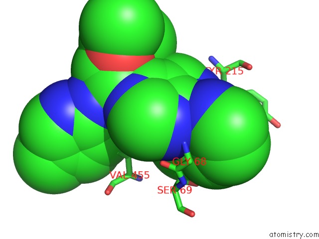
Mono view
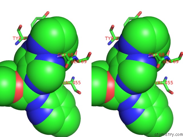
Stereo pair view
Mono view
Stereo pair view
A full contact list of Chlorine with other atoms in the Cl binding
site number 1 of Crystal Structure of Human Glucokinase in Complex with A Small Molecule Activator. within 5.0Å range:
|
Reference:
M.J.Waring,
S.N.L.Bennett,
S.Boyd,
L.Campbell,
R.D.M.Davies,
S.Gerhardt,
D.Hargreaves,
N.G Martin,
G.R.Robb,
G.Wilkinson.
Matched Triplicate Design Sets in the Optimisation of Glucokinase Activators Maximising Medicinal Chemistry Information Content To Be Published.
Page generated: Sun Jul 21 17:05:48 2024
Last articles
Ca in 5MFACa in 5MF4
Ca in 5MEY
Ca in 5MB1
Ca in 5MAZ
Ca in 5MER
Ca in 5MAY
Ca in 5MEH
Ca in 5MC9
Ca in 5MA7