Atomistry » Chlorine » PDB 4jk9-4jtg » 4jra
Atomistry »
  Chlorine »
    PDB 4jk9-4jtg »
      4jra »

Chlorine in PDB 4jra: Crystal Structure of the Botulinum Neurotoxin A Receptor-Binding Domain in Complex with the Luminal Domain of SV2C

Enzymatic activity of Crystal Structure of the Botulinum Neurotoxin A Receptor-Binding Domain in Complex with the Luminal Domain of SV2C

All present enzymatic activity of Crystal Structure of the Botulinum Neurotoxin A Receptor-Binding Domain in Complex with the Luminal Domain of SV2C:
3.4.24.69;

Protein crystallography data

The structure of Crystal Structure of the Botulinum Neurotoxin A Receptor-Binding Domain in Complex with the Luminal Domain of SV2C, PDB code: 4jra was solved by R.M.Benoit, D.Frey, M.M.Wieser, R.Jaussi, G.F.X.Schertler, G.Capitani, R.A.Kammerer, with X-Ray Crystallography technique. A brief refinement statistics is given in the table below:

Resolution Low / High (Å) 19.93 / 2.30
Space group C 1 2 1
Cell size a, b, c (Å), α, β, γ (°) 115.440, 105.260, 127.960, 90.00, 90.02, 90.00
R / Rfree (%) 23.5 / 26.9

Other elements in 4jra:

The structure of Crystal Structure of the Botulinum Neurotoxin A Receptor-Binding Domain in Complex with the Luminal Domain of SV2C also contains other interesting chemical elements:

Sodium (Na) 2 atoms

Chlorine Binding Sites:

The binding sites of Chlorine atom in the Crystal Structure of the Botulinum Neurotoxin A Receptor-Binding Domain in Complex with the Luminal Domain of SV2C (pdb code 4jra). This binding sites where shown within 5.0 Angstroms radius around Chlorine atom.
In total 6 binding sites of Chlorine where determined in the Crystal Structure of the Botulinum Neurotoxin A Receptor-Binding Domain in Complex with the Luminal Domain of SV2C, PDB code: 4jra:
Jump to Chlorine binding site number: 1; 2; 3; 4; 5; 6;

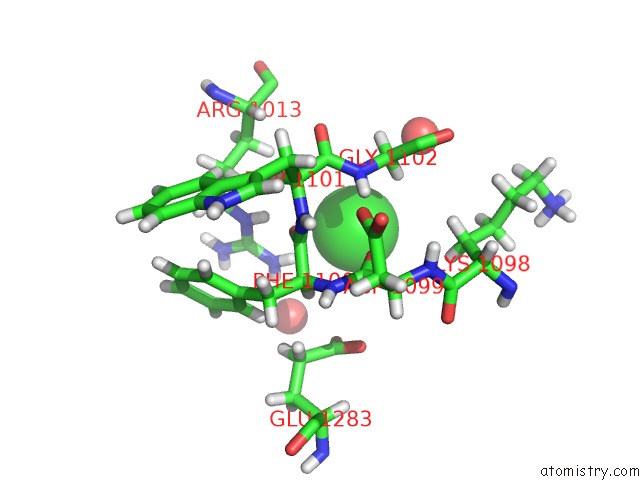
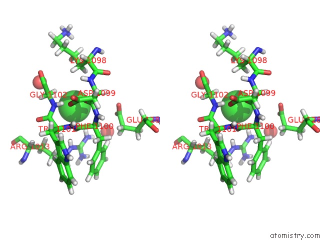
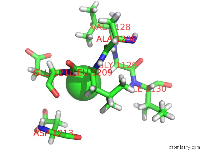
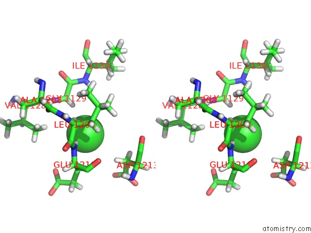
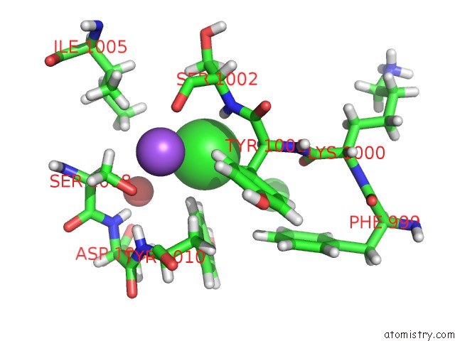
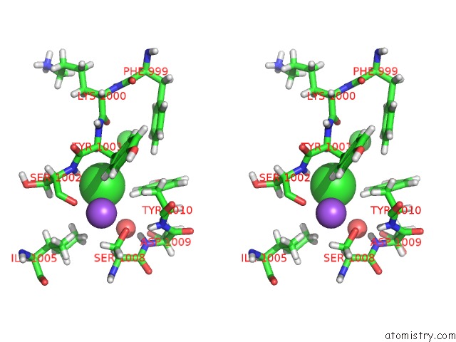
Chlorine binding site 1 out of 6 in 4jra

Go back to Chlorine Binding Sites List in 4jra
Chlorine binding site 1 out of 6 in the Crystal Structure of the Botulinum Neurotoxin A Receptor-Binding Domain in Complex with the Luminal Domain of SV2C


Mono view


Stereo pair view

A full contact list of Chlorine with other atoms in the Cl binding site number 1 of Crystal Structure of the Botulinum Neurotoxin A Receptor-Binding Domain in Complex with the Luminal Domain of SV2C within 5.0Å range:
probe atom residue distance (Å) B Occ
A:Cl1301

b:37.8
occ:1.00
HH21 A:ARG1013 2.9 16.9 1.0
O A:PHE1100 3.3 23.4 1.0
HA2 A:GLY1102 3.6 14.0 1.0
NH2 A:ARG1013 3.7 14.1 1.0
HE A:ARG1013 3.7 15.3 1.0
O A:ASP1099 3.8 11.6 1.0
C A:PHE1100 4.1 19.6 1.0
HH22 A:ARG1013 4.2 16.9 1.0
CA A:GLY1102 4.3 11.7 1.0
HA A:PHE1100 4.3 17.6 1.0
N A:GLY1102 4.3 12.1 1.0
H A:GLY1102 4.4 14.5 1.0
NE A:ARG1013 4.4 12.8 1.0
HA3 A:GLY1102 4.5 14.0 1.0
HB2 A:LYS1098 4.5 20.7 1.0
CZ A:ARG1013 4.5 11.9 1.0
O A:HOH1407 4.5 11.5 1.0
OE1 A:GLU1283 4.6 15.8 1.0
HB3 A:ARG1013 4.7 19.5 1.0
C A:TRP1101 4.7 12.9 1.0
CA A:PHE1100 4.8 14.6 1.0
HB3 A:LYS1098 4.9 20.7 1.0
C A:ASP1099 4.9 11.3 1.0
N A:TRP1101 4.9 20.3 1.0
CD A:GLU1283 5.0 18.6 1.0
O A:HOH1447 5.0 14.0 1.0

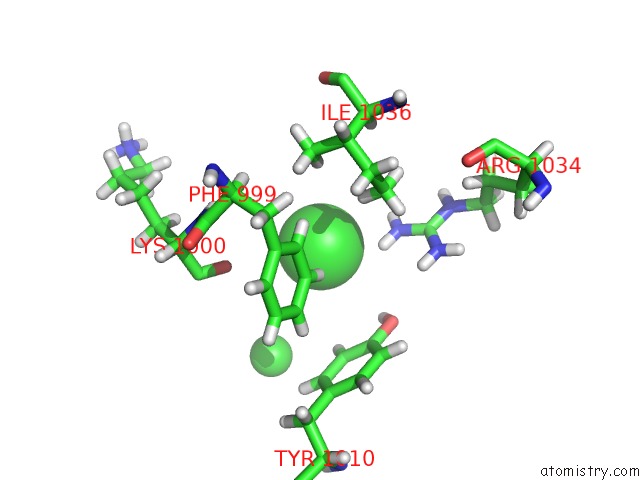
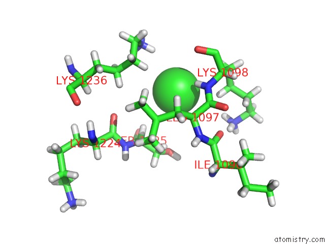
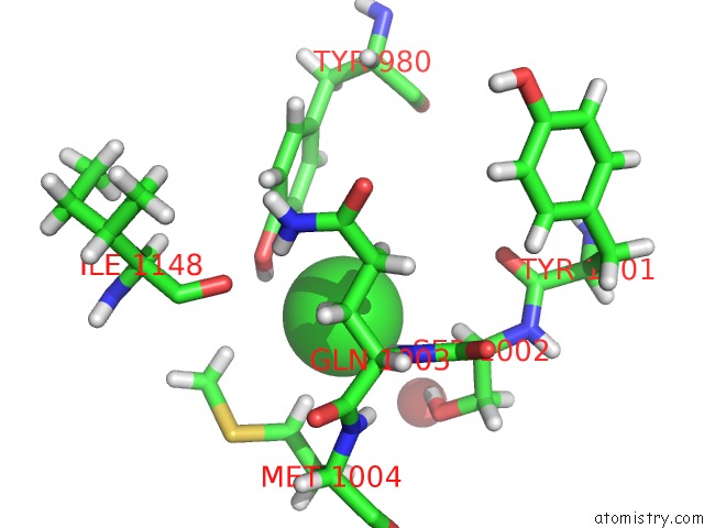
Chlorine binding site 2 out of 6 in 4jra

Go back to Chlorine Binding Sites List in 4jra
Chlorine binding site 2 out of 6 in the Crystal Structure of the Botulinum Neurotoxin A Receptor-Binding Domain in Complex with the Luminal Domain of SV2C


Mono view


Stereo pair view

A full contact list of Chlorine with other atoms in the Cl binding site number 2 of Crystal Structure of the Botulinum Neurotoxin A Receptor-Binding Domain in Complex with the Luminal Domain of SV2C within 5.0Å range:
probe atom residue distance (Å) B Occ
A:Cl1302

b:46.0
occ:1.00
HH22 A:ARG1034 2.6 50.8 1.0
HE1 A:PHE999 3.0 16.6 1.0
NH2 A:ARG1034 3.2 42.4 1.0
HH21 A:ARG1034 3.3 50.8 1.0
HD12 A:ILE1036 3.7 11.8 1.0
CE1 A:PHE999 3.8 13.8 1.0
HH A:TYR1010 3.9 17.0 1.0
HD1 A:PHE999 4.1 24.8 1.0
OH A:TYR1010 4.2 14.2 1.0
O A:LYS1000 4.2 15.0 1.0
HE2 A:TYR1010 4.2 14.2 1.0
HG23 A:ILE1036 4.3 22.1 1.0
CZ A:ARG1034 4.4 42.7 1.0
CD1 A:PHE999 4.4 20.6 1.0
HH12 A:ARG1034 4.4 53.7 1.0
CE2 A:TYR1010 4.5 11.9 1.0
CZ A:TYR1010 4.5 11.4 1.0
CD1 A:ILE1036 4.6 9.8 1.0
CZ A:PHE999 4.8 11.8 1.0
HZ A:PHE999 4.8 14.1 1.0
HD11 A:ILE1036 4.8 11.8 1.0
NH1 A:ARG1034 4.8 44.8 1.0
HD13 A:ILE1036 5.0 11.8 1.0
CL A:CL1305 5.0 32.0 1.0

Chlorine binding site 3 out of 6 in 4jra

Go back to Chlorine Binding Sites List in 4jra
Chlorine binding site 3 out of 6 in the Crystal Structure of the Botulinum Neurotoxin A Receptor-Binding Domain in Complex with the Luminal Domain of SV2C


Mono view


Stereo pair view

A full contact list of Chlorine with other atoms in the Cl binding site number 3 of Crystal Structure of the Botulinum Neurotoxin A Receptor-Binding Domain in Complex with the Luminal Domain of SV2C within 5.0Å range:
probe atom residue distance (Å) B Occ
A:Cl1303

b:30.4
occ:1.00
H A:GLU1210 2.2 31.3 1.0
HB2 A:GLU1210 2.7 25.1 1.0
HA2 A:GLY1129 2.9 34.9 1.0
N A:GLU1210 3.0 26.1 1.0
HB3 A:GLU1210 3.3 25.1 1.0
HA A:LEU1209 3.3 32.7 1.0
CB A:GLU1210 3.3 20.9 1.0
HD13 A:LEU1209 3.4 44.2 1.0
CA A:GLY1129 3.8 29.1 1.0
CA A:GLU1210 3.8 25.2 1.0
HG12 A:VAL1128 3.8 18.6 1.0
HA3 A:GLY1129 3.8 34.9 1.0
HB2 A:ASP1213 3.8 37.3 1.0
C A:LEU1209 4.0 27.0 1.0
HB3 A:LEU1209 4.0 33.3 1.0
CA A:LEU1209 4.0 27.2 1.0
HD12 A:LEU1209 4.1 44.2 1.0
CD1 A:LEU1209 4.2 36.8 1.0
N A:GLY1129 4.4 28.4 1.0
H A:ILE1130 4.4 24.3 1.0
O A:GLU1210 4.4 21.0 1.0
H A:GLY1129 4.4 34.1 1.0
CB A:LEU1209 4.4 27.7 1.0
HB3 A:ASP1213 4.5 37.3 1.0
OE1 A:GLU1210 4.5 19.9 1.0
HA A:GLU1210 4.5 30.3 1.0
CG1 A:VAL1128 4.6 15.5 1.0
CB A:ASP1213 4.6 31.1 1.0
C A:GLU1210 4.6 21.2 1.0
HG13 A:VAL1128 4.7 18.6 1.0
CG A:GLU1210 4.7 22.5 1.0
HG11 A:VAL1128 4.7 18.6 1.0
HD11 A:LEU1209 4.9 44.2 1.0
CG A:LEU1209 4.9 28.9 1.0
OD2 A:ASP1213 4.9 43.9 1.0
C A:GLY1129 4.9 24.0 1.0
HG2 A:GLU1210 4.9 27.0 1.0
O A:ALA1208 5.0 15.6 1.0
N A:ILE1130 5.0 20.3 1.0

Chlorine binding site 4 out of 6 in 4jra

Go back to Chlorine Binding Sites List in 4jra
Chlorine binding site 4 out of 6 in the Crystal Structure of the Botulinum Neurotoxin A Receptor-Binding Domain in Complex with the Luminal Domain of SV2C


Mono view


Stereo pair view

A full contact list of Chlorine with other atoms in the Cl binding site number 4 of Crystal Structure of the Botulinum Neurotoxin A Receptor-Binding Domain in Complex with the Luminal Domain of SV2C within 5.0Å range:
probe atom residue distance (Å) B Occ
A:Cl1304

b:24.1
occ:1.00
H A:LYS1098 2.5 24.3 1.0
HA A:LEU1097 2.8 21.4 1.0
HG2 A:LYS1098 3.2 14.6 1.0
N A:LYS1098 3.3 20.2 1.0
HZ2 A:LYS1236 3.5 21.9 1.0
HA A:SER1225 3.6 11.3 1.0
HD3 A:LYS1236 3.6 19.9 1.0
CA A:LEU1097 3.7 17.9 1.0
HB2 A:LYS1098 3.8 20.7 1.0
HG A:LEU1097 3.8 15.9 1.0
C A:LEU1097 3.9 19.9 1.0
CG A:LYS1098 4.0 12.2 1.0
HD23 A:LEU1097 4.2 15.2 1.0
CB A:LYS1098 4.2 17.2 1.0
HB3 A:LEU1097 4.2 18.4 1.0
HG3 A:LYS1098 4.2 14.6 1.0
HZ3 A:LYS1098 4.2 14.8 1.0
HB2 A:SER1225 4.2 12.4 1.0
CA A:LYS1098 4.2 17.4 1.0
NZ A:LYS1236 4.3 18.2 1.0
CB A:LEU1097 4.3 15.3 1.0
HZ3 A:LYS1236 4.3 21.9 1.0
O A:LYS1098 4.3 18.6 1.0
CG A:LEU1097 4.4 13.2 1.0
CA A:SER1225 4.5 9.4 1.0
CD A:LYS1236 4.5 16.6 1.0
HG2 A:LYS1236 4.5 20.1 1.0
OG A:SER1225 4.6 25.6 1.0
O A:ILE1096 4.6 26.9 1.0
CB A:SER1225 4.6 10.3 1.0
N A:LEU1097 4.8 24.2 1.0
C A:LYS1098 4.8 18.0 1.0
CD2 A:LEU1097 4.8 12.7 1.0
O A:SER1225 4.8 37.6 1.0
HZ2 A:LYS1098 4.8 14.8 1.0
HG3 A:LYS1236 4.9 20.1 1.0
CG A:LYS1236 4.9 16.8 1.0
HZ1 A:LYS1236 4.9 21.9 1.0
NZ A:LYS1098 4.9 12.3 1.0
CE A:LYS1236 5.0 20.7 1.0
HG A:SER1225 5.0 30.7 1.0
O A:LYS1224 5.0 11.7 1.0

Chlorine binding site 5 out of 6 in 4jra

Go back to Chlorine Binding Sites List in 4jra
Chlorine binding site 5 out of 6 in the Crystal Structure of the Botulinum Neurotoxin A Receptor-Binding Domain in Complex with the Luminal Domain of SV2C


Mono view


Stereo pair view

A full contact list of Chlorine with other atoms in the Cl binding site number 5 of Crystal Structure of the Botulinum Neurotoxin A Receptor-Binding Domain in Complex with the Luminal Domain of SV2C within 5.0Å range:
probe atom residue distance (Å) B Occ
A:Cl1305

b:32.0
occ:1.00
HD2 A:TYR1010 2.6 15.4 1.0
H A:SER1002 2.8 20.7 1.0
HE2 A:TYR1010 2.9 14.2 1.0
HB2 A:TYR1001 3.1 15.3 1.0
NA A:NA1306 3.2 22.3 1.0
HA A:TYR1001 3.2 19.1 1.0
CD2 A:TYR1010 3.2 12.8 1.0
CE2 A:TYR1010 3.4 11.9 1.0
N A:SER1002 3.6 17.2 1.0
CB A:TYR1001 3.8 12.7 1.0
CA A:TYR1001 3.9 15.9 1.0
HB3 A:TYR1001 3.9 15.3 1.0
O A:HOH1414 3.9 26.6 1.0
HD12 A:ILE1005 3.9 15.4 1.0
HD13 A:ILE1005 4.1 15.4 1.0
HB3 A:SER1002 4.2 25.9 1.0
C A:TYR1001 4.3 16.0 1.0
HB2 A:SER1002 4.3 25.9 1.0
CD1 A:ILE1005 4.4 12.8 1.0
CG A:TYR1010 4.5 13.5 1.0
HB2 A:TYR1010 4.5 22.4 1.0
CB A:SER1002 4.6 21.6 1.0
CA A:SER1002 4.6 18.4 1.0
O A:SER1002 4.6 19.6 1.0
CZ A:TYR1010 4.6 11.4 1.0
HD11 A:ILE1005 4.7 15.4 1.0
OG A:SER1008 4.7 12.5 1.0
H A:ASP1009 4.8 21.9 1.0
HZ A:PHE999 4.8 14.1 1.0
O A:LYS1000 4.8 15.0 1.0
HG21 A:ILE1005 4.9 15.6 1.0
HH A:TYR1010 4.9 17.0 1.0
CL A:CL1302 5.0 46.0 1.0

Chlorine binding site 6 out of 6 in 4jra

Go back to Chlorine Binding Sites List in 4jra
Chlorine binding site 6 out of 6 in the Crystal Structure of the Botulinum Neurotoxin A Receptor-Binding Domain in Complex with the Luminal Domain of SV2C


Mono view


Stereo pair view

A full contact list of Chlorine with other atoms in the Cl binding site number 6 of Crystal Structure of the Botulinum Neurotoxin A Receptor-Binding Domain in Complex with the Luminal Domain of SV2C within 5.0Å range:
probe atom residue distance (Å) B Occ
A:Cl1308

b:24.1
occ:1.00
H A:GLN1003 2.5 31.9 1.0
HH A:TYR980 2.6 25.5 1.0
HG2 A:MET1004 2.8 15.8 1.0
OH A:TYR980 3.0 21.3 1.0
HB2 A:GLN1003 3.1 29.1 1.0
N A:GLN1003 3.3 26.6 1.0
HE2 A:TYR980 3.3 19.9 1.0
H A:MET1004 3.4 30.0 1.0
HA A:SER1002 3.4 22.0 1.0
HG A:SER1002 3.5 32.4 1.0
CZ A:TYR980 3.6 21.0 1.0
HE2 A:MET1004 3.6 15.6 1.0
CG A:MET1004 3.7 13.2 1.0
HG3 A:GLN1003 3.7 23.0 1.0
CE2 A:TYR980 3.7 16.6 1.0
HG3 A:MET1004 3.8 15.8 1.0
CB A:GLN1003 3.8 24.2 1.0
N A:MET1004 3.9 25.0 1.0
OG A:SER1002 4.0 27.0 1.0
CA A:GLN1003 4.0 21.0 1.0
O A:HOH1423 4.2 10.8 1.0
CA A:SER1002 4.2 18.4 1.0
C A:SER1002 4.2 23.4 1.0
CG A:GLN1003 4.3 19.1 1.0
C A:GLN1003 4.3 22.4 1.0
CE A:MET1004 4.3 13.0 1.0
O A:ILE1148 4.4 14.6 1.0
HE3 A:MET1004 4.5 15.6 1.0
HB2 A:MET1004 4.5 10.6 1.0
CB A:MET1004 4.6 8.8 1.0
HE21 A:GLN1003 4.6 15.3 1.0
CE1 A:TYR980 4.6 18.0 1.0
HB3 A:GLN1003 4.6 29.1 1.0
SD A:MET1004 4.7 22.1 1.0
CB A:SER1002 4.7 21.6 1.0
CD2 A:TYR980 4.8 13.9 1.0
HE1 A:TYR980 4.9 21.6 1.0
HG2 A:GLN1003 4.9 23.0 1.0
CA A:MET1004 4.9 17.6 1.0
HA A:GLN1003 4.9 25.2 1.0
O A:TYR1001 5.0 18.7 1.0

Reference:

R.M.Benoit, D.Frey, M.Hilbert, J.T.Kevenaar, M.M.Wieser, C.U.Stirnimann, D.Mcmillan, T.Ceska, F.Lebon, R.Jaussi, M.O.Steinmetz, G.F.Schertler, C.C.Hoogenraad, G.Capitani, R.A.Kammerer. Structural Basis For Recognition of Synaptic Vesicle Protein 2C By Botulinum Neurotoxin A. Nature V. 505 108 2014.
ISSN: ISSN 0028-0836
PubMed: 24240280
DOI: 10.1038/NATURE12732
Page generated: Sat Dec 12 10:48:45 2020

Last articles

Zn in 8WB0
Zn in 8WAX
Zn in 8WAU
Zn in 8WAZ
Zn in 8WAY
Zn in 8WAV
Zn in 8WAW
Zn in 8WAT
Zn in 8W7M
Zn in 8WD3
© Copyright 2008-2020 by atomistry.com
Home   |    Site Map   |    Copyright   |    Contact us   |    Privacy