Chlorine »
PDB 4k2f-4kbb »
4k7v »
Chlorine in PDB 4k7v: OYE1-W116A Complexed with (R)-Carvone
Enzymatic activity of OYE1-W116A Complexed with (R)-Carvone
All present enzymatic activity of OYE1-W116A Complexed with (R)-Carvone:
1.6.99.1;
1.6.99.1;
Protein crystallography data
The structure of OYE1-W116A Complexed with (R)-Carvone, PDB code: 4k7v
was solved by
B.Sullivan,
Y.A.Pompeu,
J.D.Stewart,
with X-Ray Crystallography technique. A brief refinement statistics is given in the table below:
| Resolution Low / High (Å) | 35.42 / 1.52 |
| Space group | P 43 21 2 |
| Cell size a, b, c (Å), α, β, γ (°) | 140.853, 140.853, 42.841, 90.00, 90.00, 90.00 |
| R / Rfree (%) | 14.5 / 18.3 |
Other elements in 4k7v:
The structure of OYE1-W116A Complexed with (R)-Carvone also contains other interesting chemical elements:
| Magnesium | (Mg) | 1 atom |
| Sodium | (Na) | 2 atoms |
Chlorine Binding Sites:
The binding sites of Chlorine atom in the OYE1-W116A Complexed with (R)-Carvone
(pdb code 4k7v). This binding sites where shown within
5.0 Angstroms radius around Chlorine atom.
In total only one binding site of Chlorine was determined in the OYE1-W116A Complexed with (R)-Carvone, PDB code: 4k7v:
In total only one binding site of Chlorine was determined in the OYE1-W116A Complexed with (R)-Carvone, PDB code: 4k7v:
Chlorine binding site 1 out of 1 in 4k7v
Go back to
Chlorine binding site 1 out
of 1 in the OYE1-W116A Complexed with (R)-Carvone
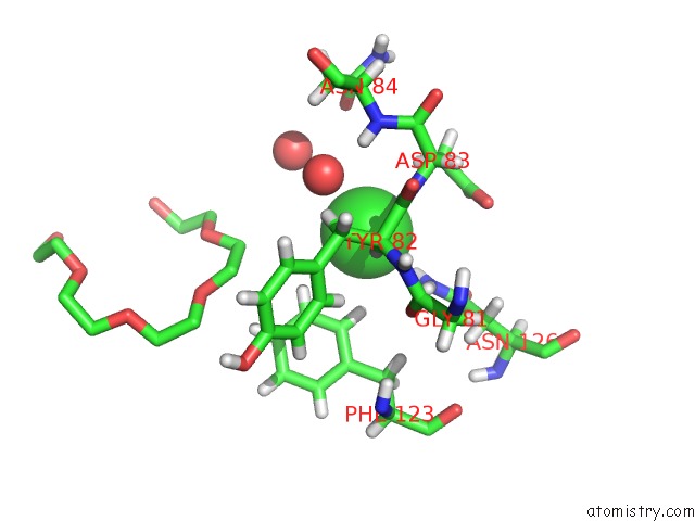
Mono view
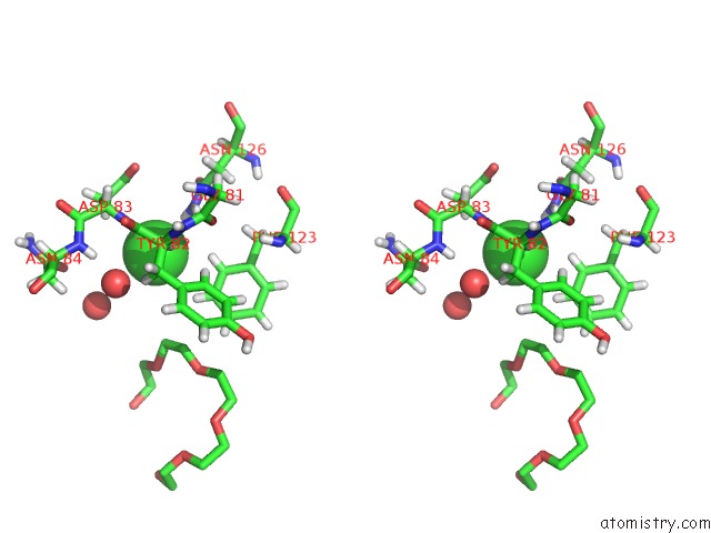
Stereo pair view
Mono view
Stereo pair view
A full contact list of Chlorine with other atoms in the Cl binding
site number 1 of OYE1-W116A Complexed with (R)-Carvone within 5.0Å range:
|
Reference:
Y.A.Pompeu,
B.Sullivan,
J.D.Stewart.
X‑Ray Crystallography Reveals How Subtle Changes Control the Orientation of Substrate Binding in An Alkene Reductase Acs Catalysis V. 3 2376 2013.
ISSN: ESSN 2155-5435
DOI: 10.1021/CS400622E
Page generated: Fri Jul 11 17:41:19 2025
ISSN: ESSN 2155-5435
DOI: 10.1021/CS400622E
Last articles
F in 7RRBF in 7RPZ
F in 7ROT
F in 7ROU
F in 7ROS
F in 7RMZ
F in 7RN6
F in 7RMJ
F in 7RJ4
F in 7RJ2