Chlorine »
PDB 4kn0-4kwt »
4kqo »
Chlorine in PDB 4kqo: Crystal Structure of Penicillin-Binding Protein 3 From Pseudomonas Aeruginosa in Complex with Piperacillin
Protein crystallography data
The structure of Crystal Structure of Penicillin-Binding Protein 3 From Pseudomonas Aeruginosa in Complex with Piperacillin, PDB code: 4kqo
was solved by
J.E.Nettleship,
D.I.Stuart,
R.J.Owens,
J.Ren,
with X-Ray Crystallography technique. A brief refinement statistics is given in the table below:
| Resolution Low / High (Å) | 47.10 / 2.31 |
| Space group | P 1 |
| Cell size a, b, c (Å), α, β, γ (°) | 57.049, 74.935, 82.785, 71.26, 86.04, 85.59 |
| R / Rfree (%) | 21.7 / 25 |
Chlorine Binding Sites:
The binding sites of Chlorine atom in the Crystal Structure of Penicillin-Binding Protein 3 From Pseudomonas Aeruginosa in Complex with Piperacillin
(pdb code 4kqo). This binding sites where shown within
5.0 Angstroms radius around Chlorine atom.
In total 2 binding sites of Chlorine where determined in the Crystal Structure of Penicillin-Binding Protein 3 From Pseudomonas Aeruginosa in Complex with Piperacillin, PDB code: 4kqo:
Jump to Chlorine binding site number: 1; 2;
In total 2 binding sites of Chlorine where determined in the Crystal Structure of Penicillin-Binding Protein 3 From Pseudomonas Aeruginosa in Complex with Piperacillin, PDB code: 4kqo:
Jump to Chlorine binding site number: 1; 2;
Chlorine binding site 1 out of 2 in 4kqo
Go back to
Chlorine binding site 1 out
of 2 in the Crystal Structure of Penicillin-Binding Protein 3 From Pseudomonas Aeruginosa in Complex with Piperacillin
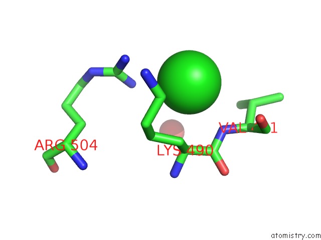
Mono view
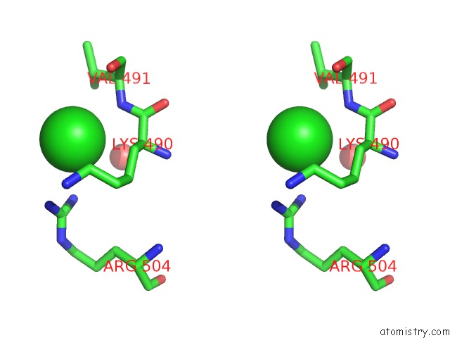
Stereo pair view
Mono view
Stereo pair view
A full contact list of Chlorine with other atoms in the Cl binding
site number 1 of Crystal Structure of Penicillin-Binding Protein 3 From Pseudomonas Aeruginosa in Complex with Piperacillin within 5.0Å range:
|
Chlorine binding site 2 out of 2 in 4kqo
Go back to
Chlorine binding site 2 out
of 2 in the Crystal Structure of Penicillin-Binding Protein 3 From Pseudomonas Aeruginosa in Complex with Piperacillin
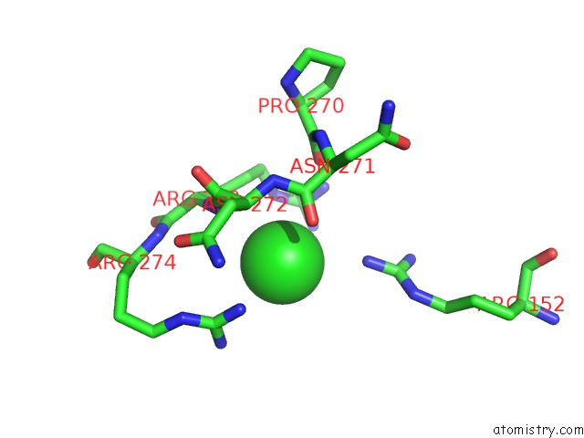
Mono view
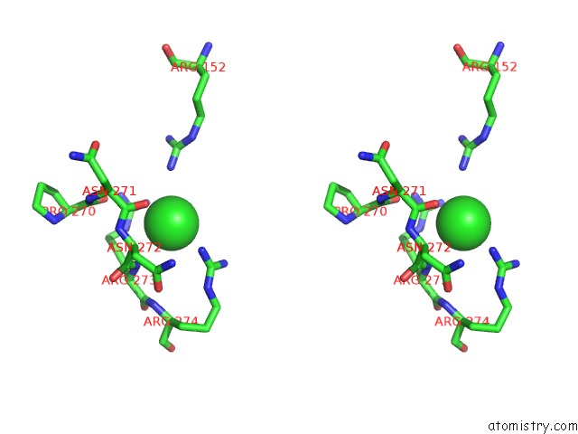
Stereo pair view
Mono view
Stereo pair view
A full contact list of Chlorine with other atoms in the Cl binding
site number 2 of Crystal Structure of Penicillin-Binding Protein 3 From Pseudomonas Aeruginosa in Complex with Piperacillin within 5.0Å range:
|
Reference:
S.S.Van Berkel,
J.E.Nettleship,
I.K.Leung,
J.Brem,
H.Choi,
D.I.Stuart,
T.D.Claridge,
M.A.Mcdonough,
R.J.Owens,
J.Ren,
C.J.Schofield.
Binding of (5S)-Penicilloic Acid to Penicillin Binding Protein 3. Acs Chem.Biol. V. 8 2112 2013.
ISSN: ISSN 1554-8929
PubMed: 23899657
DOI: 10.1021/CB400200H
Page generated: Fri Jul 11 17:53:22 2025
ISSN: ISSN 1554-8929
PubMed: 23899657
DOI: 10.1021/CB400200H
Last articles
F in 4KHRF in 4KIL
F in 4KGJ
F in 4KFO
F in 4KH2
F in 4KBK
F in 4KFG
F in 4KCS
F in 4KCR
F in 4KCN