Chlorine »
PDB 4kn0-4kwt »
4ksi »
Chlorine in PDB 4ksi: Crystal Structure Analysis of the Acidic Leucine Aminopeptidase of Tomato
Enzymatic activity of Crystal Structure Analysis of the Acidic Leucine Aminopeptidase of Tomato
All present enzymatic activity of Crystal Structure Analysis of the Acidic Leucine Aminopeptidase of Tomato:
3.4.11.1; 3.4.11.5;
3.4.11.1; 3.4.11.5;
Protein crystallography data
The structure of Crystal Structure Analysis of the Acidic Leucine Aminopeptidase of Tomato, PDB code: 4ksi
was solved by
K.T.Duprez,
M.Scranton,
L.Walling,
L.Fan,
with X-Ray Crystallography technique. A brief refinement statistics is given in the table below:
| Resolution Low / High (Å) | 29.14 / 2.20 |
| Space group | P 63 2 2 |
| Cell size a, b, c (Å), α, β, γ (°) | 160.597, 160.597, 104.183, 90.00, 90.00, 120.00 |
| R / Rfree (%) | 13.7 / 17.4 |
Other elements in 4ksi:
The structure of Crystal Structure Analysis of the Acidic Leucine Aminopeptidase of Tomato also contains other interesting chemical elements:
| Magnesium | (Mg) | 2 atoms |
Chlorine Binding Sites:
The binding sites of Chlorine atom in the Crystal Structure Analysis of the Acidic Leucine Aminopeptidase of Tomato
(pdb code 4ksi). This binding sites where shown within
5.0 Angstroms radius around Chlorine atom.
In total 3 binding sites of Chlorine where determined in the Crystal Structure Analysis of the Acidic Leucine Aminopeptidase of Tomato, PDB code: 4ksi:
Jump to Chlorine binding site number: 1; 2; 3;
In total 3 binding sites of Chlorine where determined in the Crystal Structure Analysis of the Acidic Leucine Aminopeptidase of Tomato, PDB code: 4ksi:
Jump to Chlorine binding site number: 1; 2; 3;
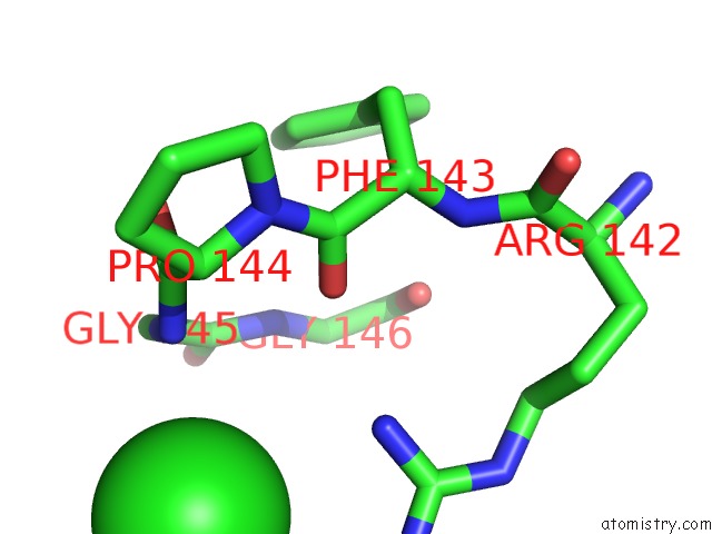
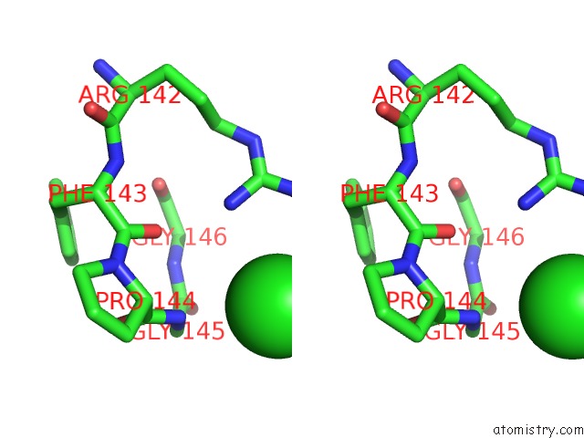
Chlorine binding site 1 out of 3 in 4ksi
Go back to
Chlorine binding site 1 out
of 3 in the Crystal Structure Analysis of the Acidic Leucine Aminopeptidase of Tomato
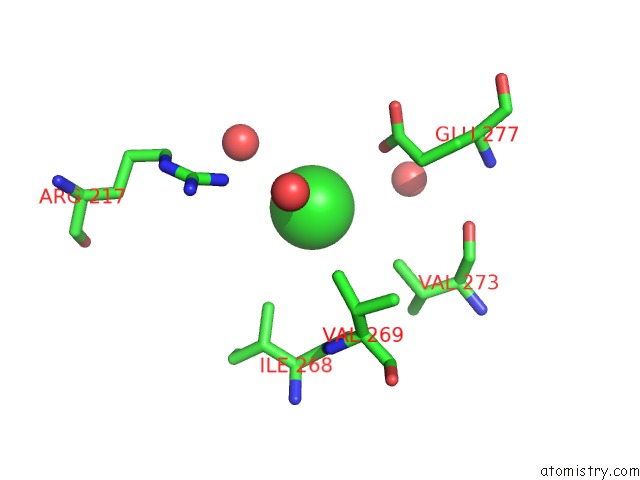
Mono view
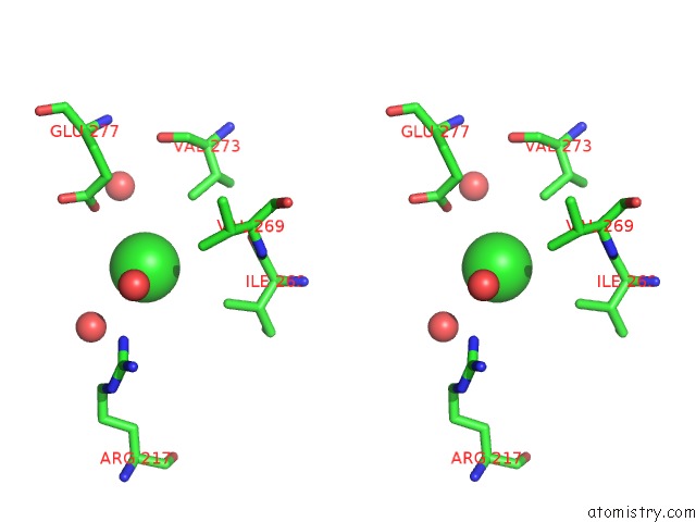
Stereo pair view
Mono view
Stereo pair view
A full contact list of Chlorine with other atoms in the Cl binding
site number 1 of Crystal Structure Analysis of the Acidic Leucine Aminopeptidase of Tomato within 5.0Å range:
|
Chlorine binding site 2 out of 3 in 4ksi
Go back to
Chlorine binding site 2 out
of 3 in the Crystal Structure Analysis of the Acidic Leucine Aminopeptidase of Tomato
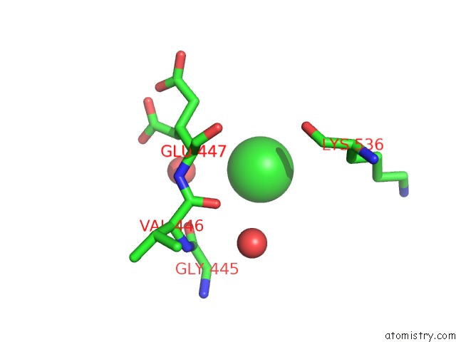
Mono view
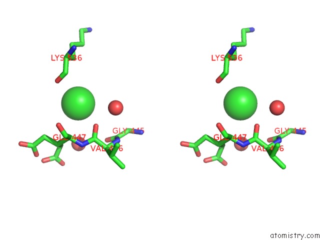
Stereo pair view
Mono view
Stereo pair view
A full contact list of Chlorine with other atoms in the Cl binding
site number 2 of Crystal Structure Analysis of the Acidic Leucine Aminopeptidase of Tomato within 5.0Å range:
|
Chlorine binding site 3 out of 3 in 4ksi
Go back to
Chlorine binding site 3 out
of 3 in the Crystal Structure Analysis of the Acidic Leucine Aminopeptidase of Tomato
Mono view
Stereo pair view
Mono view
Stereo pair view
A full contact list of Chlorine with other atoms in the Cl binding
site number 3 of Crystal Structure Analysis of the Acidic Leucine Aminopeptidase of Tomato within 5.0Å range:
|
Reference:
K.Duprez,
M.A.Scranton,
L.L.Walling,
L.Fan.
Structure of Tomato Wound-Induced Leucine Aminopeptidase Sheds Light on Substrate Specificity. Acta Crystallogr.,Sect.D V. 70 1649 2014.
ISSN: ISSN 0907-4449
PubMed: 24914976
DOI: 10.1107/S1399004714006245
Page generated: Sun Jul 21 18:23:36 2024
ISSN: ISSN 0907-4449
PubMed: 24914976
DOI: 10.1107/S1399004714006245
Last articles
Zn in 9MJ5Zn in 9HNW
Zn in 9G0L
Zn in 9FNE
Zn in 9DZN
Zn in 9E0I
Zn in 9D32
Zn in 9DAK
Zn in 8ZXC
Zn in 8ZUF