Chlorine »
PDB 4lbb-4lhu »
4lgl »
Chlorine in PDB 4lgl: Crystal Structure of Glycine Decarboxylase P-Protein From Synechocystis Sp. Pcc 6803, Apo Form
Enzymatic activity of Crystal Structure of Glycine Decarboxylase P-Protein From Synechocystis Sp. Pcc 6803, Apo Form
All present enzymatic activity of Crystal Structure of Glycine Decarboxylase P-Protein From Synechocystis Sp. Pcc 6803, Apo Form:
1.4.4.2;
1.4.4.2;
Protein crystallography data
The structure of Crystal Structure of Glycine Decarboxylase P-Protein From Synechocystis Sp. Pcc 6803, Apo Form, PDB code: 4lgl
was solved by
D.Hasse,
E.Andersson,
G.Carlsson,
A.Masloboy,
M.Hagemann,
H.Bauwe,
I.Andersson,
with X-Ray Crystallography technique. A brief refinement statistics is given in the table below:
| Resolution Low / High (Å) | 54.11 / 2.00 |
| Space group | P 21 21 21 |
| Cell size a, b, c (Å), α, β, γ (°) | 96.299, 135.814, 179.080, 90.00, 90.00, 90.00 |
| R / Rfree (%) | 16.1 / 19.2 |
Chlorine Binding Sites:
The binding sites of Chlorine atom in the Crystal Structure of Glycine Decarboxylase P-Protein From Synechocystis Sp. Pcc 6803, Apo Form
(pdb code 4lgl). This binding sites where shown within
5.0 Angstroms radius around Chlorine atom.
In total 2 binding sites of Chlorine where determined in the Crystal Structure of Glycine Decarboxylase P-Protein From Synechocystis Sp. Pcc 6803, Apo Form, PDB code: 4lgl:
Jump to Chlorine binding site number: 1; 2;
In total 2 binding sites of Chlorine where determined in the Crystal Structure of Glycine Decarboxylase P-Protein From Synechocystis Sp. Pcc 6803, Apo Form, PDB code: 4lgl:
Jump to Chlorine binding site number: 1; 2;
Chlorine binding site 1 out of 2 in 4lgl
Go back to
Chlorine binding site 1 out
of 2 in the Crystal Structure of Glycine Decarboxylase P-Protein From Synechocystis Sp. Pcc 6803, Apo Form
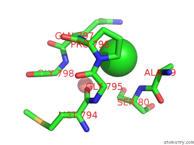
Mono view
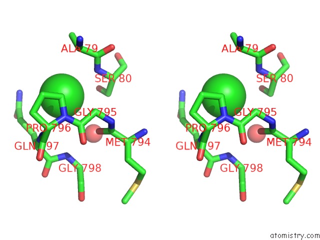
Stereo pair view
Mono view
Stereo pair view
A full contact list of Chlorine with other atoms in the Cl binding
site number 1 of Crystal Structure of Glycine Decarboxylase P-Protein From Synechocystis Sp. Pcc 6803, Apo Form within 5.0Å range:
|
Chlorine binding site 2 out of 2 in 4lgl
Go back to
Chlorine binding site 2 out
of 2 in the Crystal Structure of Glycine Decarboxylase P-Protein From Synechocystis Sp. Pcc 6803, Apo Form
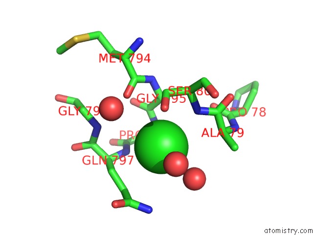
Mono view
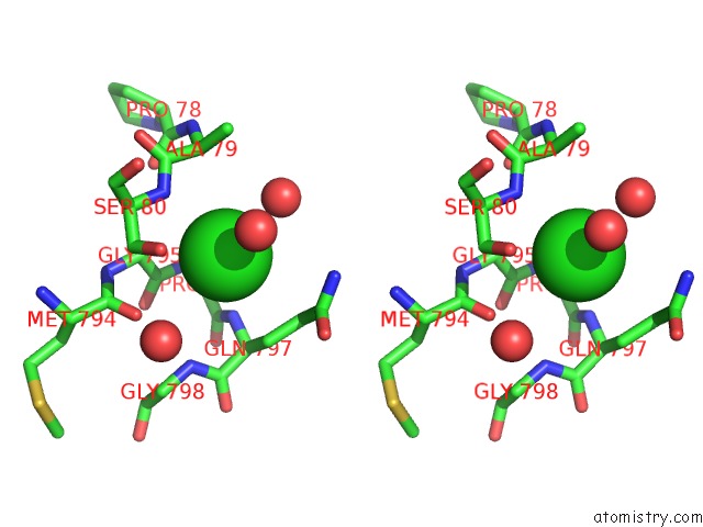
Stereo pair view
Mono view
Stereo pair view
A full contact list of Chlorine with other atoms in the Cl binding
site number 2 of Crystal Structure of Glycine Decarboxylase P-Protein From Synechocystis Sp. Pcc 6803, Apo Form within 5.0Å range:
|
Reference:
D.Hasse,
E.Andersson,
G.Carlsson,
A.Masloboy,
M.Hagemann,
H.Bauwe,
I.Andersson.
Structure of the Homodimeric Glycine Decarboxylase P-Protein From Synechocystis Sp. Pcc 6803 Suggests A Mechanism For Redox Regulation. J.Biol.Chem. V. 288 35333 2013.
ISSN: ISSN 0021-9258
PubMed: 24121504
DOI: 10.1074/JBC.M113.509976
Page generated: Sun Jul 21 18:55:02 2024
ISSN: ISSN 0021-9258
PubMed: 24121504
DOI: 10.1074/JBC.M113.509976
Last articles
Zn in 9J0NZn in 9J0O
Zn in 9J0P
Zn in 9FJX
Zn in 9EKB
Zn in 9C0F
Zn in 9CAH
Zn in 9CH0
Zn in 9CH3
Zn in 9CH1