Chlorine »
PDB 4li5-4lqz »
4lpq »
Chlorine in PDB 4lpq: Crystal Structure of the L,D-Transpeptidase (Residues 123-326) From Xylanimonas Cellulosilytica Dsm 15894
Protein crystallography data
The structure of Crystal Structure of the L,D-Transpeptidase (Residues 123-326) From Xylanimonas Cellulosilytica Dsm 15894, PDB code: 4lpq
was solved by
B.Nocek,
L.Bigelow,
M.Endres,
G.Babnigg,
A.Joachimiak,
Midwest Center Forstructural Genomics (Mcsg),
with X-Ray Crystallography technique. A brief refinement statistics is given in the table below:
| Resolution Low / High (Å) | 23.61 / 1.37 |
| Space group | P 43 21 2 |
| Cell size a, b, c (Å), α, β, γ (°) | 62.849, 62.849, 167.210, 90.00, 90.00, 90.00 |
| R / Rfree (%) | 12.2 / 14.6 |
Chlorine Binding Sites:
The binding sites of Chlorine atom in the Crystal Structure of the L,D-Transpeptidase (Residues 123-326) From Xylanimonas Cellulosilytica Dsm 15894
(pdb code 4lpq). This binding sites where shown within
5.0 Angstroms radius around Chlorine atom.
In total 4 binding sites of Chlorine where determined in the Crystal Structure of the L,D-Transpeptidase (Residues 123-326) From Xylanimonas Cellulosilytica Dsm 15894, PDB code: 4lpq:
Jump to Chlorine binding site number: 1; 2; 3; 4;
In total 4 binding sites of Chlorine where determined in the Crystal Structure of the L,D-Transpeptidase (Residues 123-326) From Xylanimonas Cellulosilytica Dsm 15894, PDB code: 4lpq:
Jump to Chlorine binding site number: 1; 2; 3; 4;
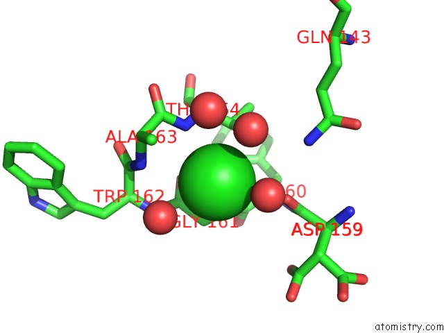
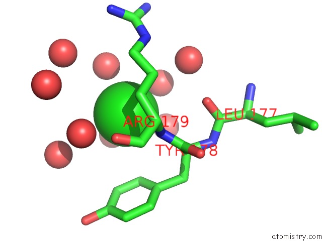
Chlorine binding site 1 out of 4 in 4lpq
Go back to
Chlorine binding site 1 out
of 4 in the Crystal Structure of the L,D-Transpeptidase (Residues 123-326) From Xylanimonas Cellulosilytica Dsm 15894
Mono view
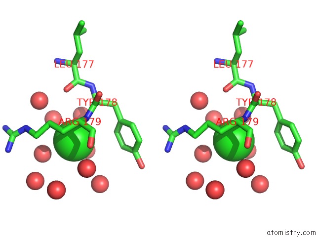
Stereo pair view
Mono view
Stereo pair view
A full contact list of Chlorine with other atoms in the Cl binding
site number 1 of Crystal Structure of the L,D-Transpeptidase (Residues 123-326) From Xylanimonas Cellulosilytica Dsm 15894 within 5.0Å range:
|
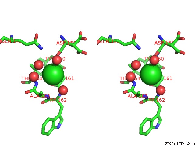
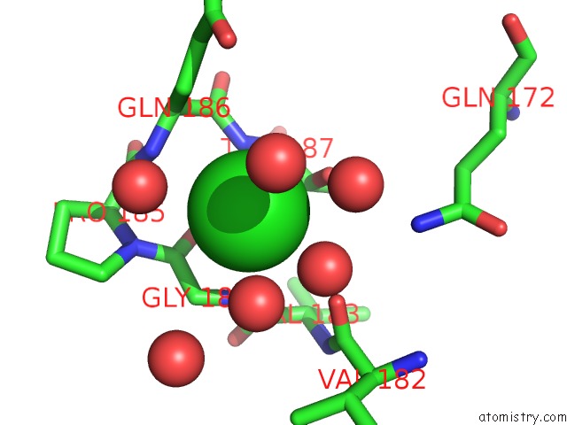
Chlorine binding site 2 out of 4 in 4lpq
Go back to
Chlorine binding site 2 out
of 4 in the Crystal Structure of the L,D-Transpeptidase (Residues 123-326) From Xylanimonas Cellulosilytica Dsm 15894
Mono view
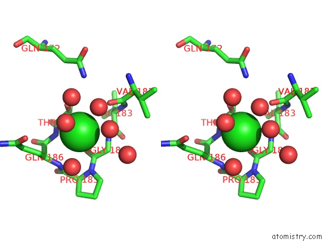
Stereo pair view
Mono view
Stereo pair view
A full contact list of Chlorine with other atoms in the Cl binding
site number 2 of Crystal Structure of the L,D-Transpeptidase (Residues 123-326) From Xylanimonas Cellulosilytica Dsm 15894 within 5.0Å range:
|
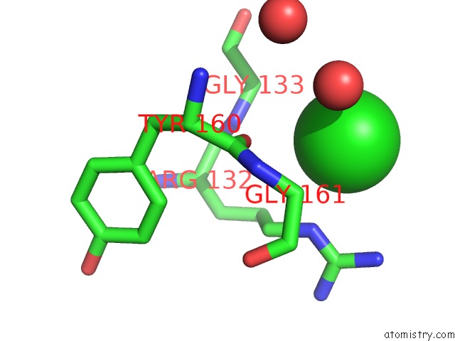
Chlorine binding site 3 out of 4 in 4lpq
Go back to
Chlorine binding site 3 out
of 4 in the Crystal Structure of the L,D-Transpeptidase (Residues 123-326) From Xylanimonas Cellulosilytica Dsm 15894
Mono view
Stereo pair view
Mono view
Stereo pair view
A full contact list of Chlorine with other atoms in the Cl binding
site number 3 of Crystal Structure of the L,D-Transpeptidase (Residues 123-326) From Xylanimonas Cellulosilytica Dsm 15894 within 5.0Å range:
|
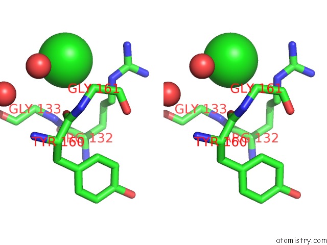
Chlorine binding site 4 out of 4 in 4lpq
Go back to
Chlorine binding site 4 out
of 4 in the Crystal Structure of the L,D-Transpeptidase (Residues 123-326) From Xylanimonas Cellulosilytica Dsm 15894
Mono view
Stereo pair view
Mono view
Stereo pair view
A full contact list of Chlorine with other atoms in the Cl binding
site number 4 of Crystal Structure of the L,D-Transpeptidase (Residues 123-326) From Xylanimonas Cellulosilytica Dsm 15894 within 5.0Å range:
|
Reference:
B.Nocek,
L.Bigelow,
M.Endres,
G.Babnigg,
A.Joachimiak.
Crystal Structure of the L,D-Transpeptidase (Residues 123-326) From Xylanimonas Cellulosilytica Dsm 15894 To Be Published.
Page generated: Fri Jul 11 18:39:55 2025
Last articles
Fe in 2YXOFe in 2YRS
Fe in 2YXC
Fe in 2YNM
Fe in 2YVJ
Fe in 2YP1
Fe in 2YU2
Fe in 2YU1
Fe in 2YQB
Fe in 2YOO