Chlorine »
PDB 4m5h-4mdq »
4ma8 »
Chlorine in PDB 4ma8: Crystal Structure of Mouse Prion Protein Complexed with Chlorpromazine
Protein crystallography data
The structure of Crystal Structure of Mouse Prion Protein Complexed with Chlorpromazine, PDB code: 4ma8
was solved by
P.K.Baral,
M.Swayampakula,
M.N.G.James,
with X-Ray Crystallography technique. A brief refinement statistics is given in the table below:
| Resolution Low / High (Å) | 34.99 / 2.20 |
| Space group | C 1 2 1 |
| Cell size a, b, c (Å), α, β, γ (°) | 83.262, 106.883, 75.594, 90.00, 95.32, 90.00 |
| R / Rfree (%) | 19.5 / 23.6 |
Chlorine Binding Sites:
The binding sites of Chlorine atom in the Crystal Structure of Mouse Prion Protein Complexed with Chlorpromazine
(pdb code 4ma8). This binding sites where shown within
5.0 Angstroms radius around Chlorine atom.
In total only one binding site of Chlorine was determined in the Crystal Structure of Mouse Prion Protein Complexed with Chlorpromazine, PDB code: 4ma8:
In total only one binding site of Chlorine was determined in the Crystal Structure of Mouse Prion Protein Complexed with Chlorpromazine, PDB code: 4ma8:
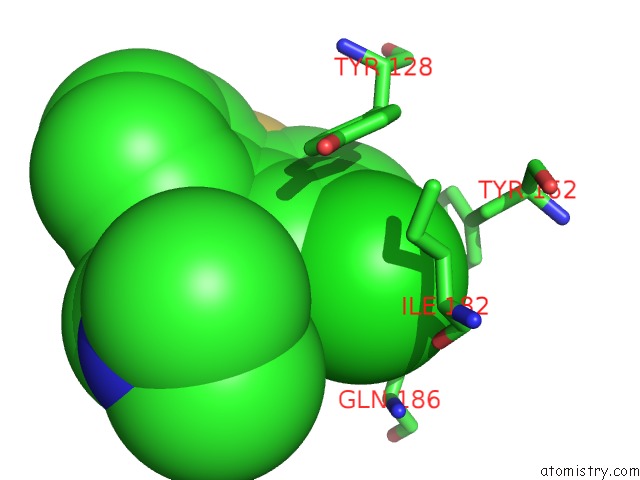
Chlorine binding site 1 out of 1 in 4ma8
Go back to
Chlorine binding site 1 out
of 1 in the Crystal Structure of Mouse Prion Protein Complexed with Chlorpromazine
Mono view

Stereo pair view
Mono view
Stereo pair view
A full contact list of Chlorine with other atoms in the Cl binding
site number 1 of Crystal Structure of Mouse Prion Protein Complexed with Chlorpromazine within 5.0Å range:
|
Reference:
P.K.Baral,
M.Swayampakula,
M.K.Rout,
N.N.Kav,
L.Spyracopoulos,
A.Aguzzi,
M.N.James.
Structural Basis of Prion Inhibition By Phenothiazine Compounds. Structure V. 22 291 2014.
ISSN: ISSN 0969-2126
PubMed: 24373770
DOI: 10.1016/J.STR.2013.11.009
Page generated: Fri Jul 11 18:58:37 2025
ISSN: ISSN 0969-2126
PubMed: 24373770
DOI: 10.1016/J.STR.2013.11.009
Last articles
Cl in 5BWHCl in 5BVZ
Cl in 5BVI
Cl in 5BW7
Cl in 5BVP
Cl in 5BVW
Cl in 5BVG
Cl in 5BVH
Cl in 5BVK
Cl in 5BVF