Chlorine »
PDB 4m5h-4mdq »
4mb3 »
Chlorine in PDB 4mb3: Crystal Structure of E153Q Mutant of Cold-Adapted Chitinase From Moritella Marina
Enzymatic activity of Crystal Structure of E153Q Mutant of Cold-Adapted Chitinase From Moritella Marina
All present enzymatic activity of Crystal Structure of E153Q Mutant of Cold-Adapted Chitinase From Moritella Marina:
3.2.1.14;
3.2.1.14;
Protein crystallography data
The structure of Crystal Structure of E153Q Mutant of Cold-Adapted Chitinase From Moritella Marina, PDB code: 4mb3
was solved by
P.H.Malecki,
C.E.Vorgias,
W.Rypniewski,
with X-Ray Crystallography technique. A brief refinement statistics is given in the table below:
| Resolution Low / High (Å) | 31.44 / 1.55 |
| Space group | P 31 1 2 |
| Cell size a, b, c (Å), α, β, γ (°) | 67.657, 67.657, 255.643, 90.00, 90.00, 120.00 |
| R / Rfree (%) | 14.1 / 18.3 |
Other elements in 4mb3:
The structure of Crystal Structure of E153Q Mutant of Cold-Adapted Chitinase From Moritella Marina also contains other interesting chemical elements:
| Sodium | (Na) | 4 atoms |
Chlorine Binding Sites:
The binding sites of Chlorine atom in the Crystal Structure of E153Q Mutant of Cold-Adapted Chitinase From Moritella Marina
(pdb code 4mb3). This binding sites where shown within
5.0 Angstroms radius around Chlorine atom.
In total only one binding site of Chlorine was determined in the Crystal Structure of E153Q Mutant of Cold-Adapted Chitinase From Moritella Marina, PDB code: 4mb3:
In total only one binding site of Chlorine was determined in the Crystal Structure of E153Q Mutant of Cold-Adapted Chitinase From Moritella Marina, PDB code: 4mb3:
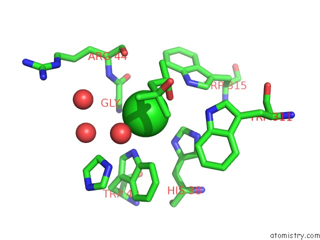
Chlorine binding site 1 out of 1 in 4mb3
Go back to
Chlorine binding site 1 out
of 1 in the Crystal Structure of E153Q Mutant of Cold-Adapted Chitinase From Moritella Marina
Mono view

Stereo pair view
Mono view
Stereo pair view
A full contact list of Chlorine with other atoms in the Cl binding
site number 1 of Crystal Structure of E153Q Mutant of Cold-Adapted Chitinase From Moritella Marina within 5.0Å range:
|
Reference:
P.H.Malecki,
C.E.Vorgias,
M.V.Petoukhov,
D.I.Svergun,
W.Rypniewski.
Crystal Structures of Substrate-Bound Chitinase From the Psychrophilic Bacterium Moritella Marina and Its Structure in Solution Acta Crystallogr.,Sect.D V. 70 676 2014.
ISSN: ISSN 0907-4449
PubMed: 24598737
DOI: 10.1107/S1399004713032264
Page generated: Fri Jul 11 18:59:15 2025
ISSN: ISSN 0907-4449
PubMed: 24598737
DOI: 10.1107/S1399004713032264
Last articles
Fe in 2YXOFe in 2YRS
Fe in 2YXC
Fe in 2YNM
Fe in 2YVJ
Fe in 2YP1
Fe in 2YU2
Fe in 2YU1
Fe in 2YQB
Fe in 2YOO