Chlorine »
PDB 4me9-4ml5 »
4miv »
Chlorine in PDB 4miv: Crystal Structure of Sulfamidase, Crystal Form L
Enzymatic activity of Crystal Structure of Sulfamidase, Crystal Form L
All present enzymatic activity of Crystal Structure of Sulfamidase, Crystal Form L:
3.10.1.1;
3.10.1.1;
Protein crystallography data
The structure of Crystal Structure of Sulfamidase, Crystal Form L, PDB code: 4miv
was solved by
N.S.Sidhu,
I.Uson,
K.Schreiber,
K.Proepper,
S.Becker,
G.M.Sheldrick,
J.Gaertner,
R.Kraetzner,
R.Steinfeld,
with X-Ray Crystallography technique. A brief refinement statistics is given in the table below:
| Resolution Low / High (Å) | 48.85 / 2.40 |
| Space group | P 1 21 1 |
| Cell size a, b, c (Å), α, β, γ (°) | 102.930, 211.450, 108.350, 90.00, 102.69, 90.00 |
| R / Rfree (%) | 21.6 / 24.5 |
Other elements in 4miv:
The structure of Crystal Structure of Sulfamidase, Crystal Form L also contains other interesting chemical elements:
| Calcium | (Ca) | 8 atoms |
Chlorine Binding Sites:
The binding sites of Chlorine atom in the Crystal Structure of Sulfamidase, Crystal Form L
(pdb code 4miv). This binding sites where shown within
5.0 Angstroms radius around Chlorine atom.
In total 2 binding sites of Chlorine where determined in the Crystal Structure of Sulfamidase, Crystal Form L, PDB code: 4miv:
Jump to Chlorine binding site number: 1; 2;
In total 2 binding sites of Chlorine where determined in the Crystal Structure of Sulfamidase, Crystal Form L, PDB code: 4miv:
Jump to Chlorine binding site number: 1; 2;
Chlorine binding site 1 out of 2 in 4miv
Go back to
Chlorine binding site 1 out
of 2 in the Crystal Structure of Sulfamidase, Crystal Form L
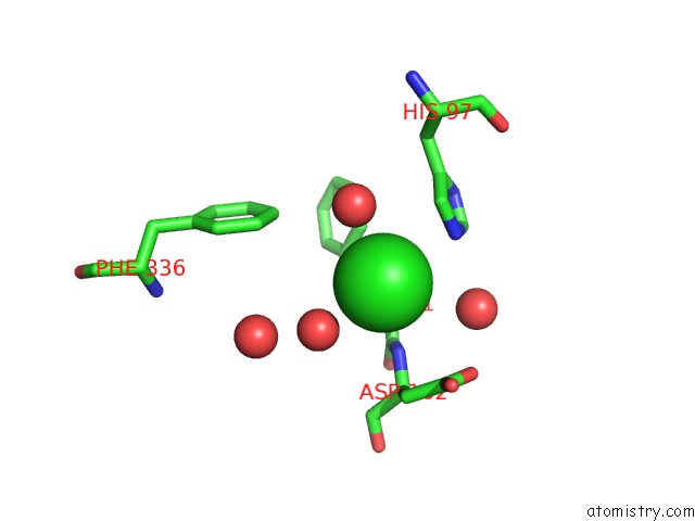
Mono view
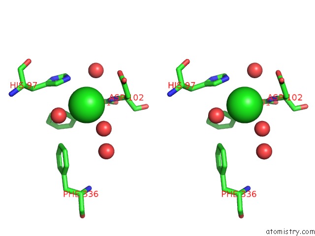
Stereo pair view
Mono view
Stereo pair view
A full contact list of Chlorine with other atoms in the Cl binding
site number 1 of Crystal Structure of Sulfamidase, Crystal Form L within 5.0Å range:
|
Chlorine binding site 2 out of 2 in 4miv
Go back to
Chlorine binding site 2 out
of 2 in the Crystal Structure of Sulfamidase, Crystal Form L
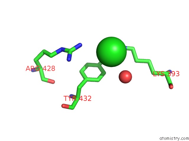
Mono view
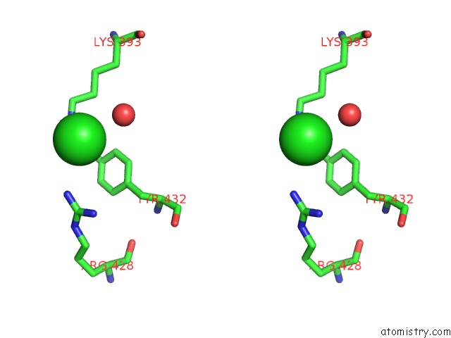
Stereo pair view
Mono view
Stereo pair view
A full contact list of Chlorine with other atoms in the Cl binding
site number 2 of Crystal Structure of Sulfamidase, Crystal Form L within 5.0Å range:
|
Reference:
N.S.Sidhu,
K.Schreiber,
K.Proepper,
S.Becker,
I.Uson,
G.M.Sheldrick,
J.Gaertner,
R.Kraetzner,
R.Steinfeld.
Structure of Sulfamidase Provides Insight Into the Molecular Pathology of Mucopolysaccharidosis Iiia Acta Crystallogr.,Sect.D V. D70 1321.
ISSN: ISSN 0907-4449
Page generated: Sun Jul 21 19:54:30 2024
ISSN: ISSN 0907-4449
Last articles
Zn in 9J0NZn in 9J0O
Zn in 9J0P
Zn in 9FJX
Zn in 9EKB
Zn in 9C0F
Zn in 9CAH
Zn in 9CH0
Zn in 9CH3
Zn in 9CH1