Chlorine »
PDB 4me9-4ml5 »
4mks »
Chlorine in PDB 4mks: Crystal Structure of Enolase From Lactobacillus Gasseri
Enzymatic activity of Crystal Structure of Enolase From Lactobacillus Gasseri
All present enzymatic activity of Crystal Structure of Enolase From Lactobacillus Gasseri:
4.2.1.11;
4.2.1.11;
Protein crystallography data
The structure of Crystal Structure of Enolase From Lactobacillus Gasseri, PDB code: 4mks
was solved by
K.Raghunathan,
P.T.Harris,
R.R.Spurbeck,
C.G.Arvidson,
D.N.Arvidson,
with X-Ray Crystallography technique. A brief refinement statistics is given in the table below:
| Resolution Low / High (Å) | 54.45 / 2.08 |
| Space group | I 4 |
| Cell size a, b, c (Å), α, β, γ (°) | 145.240, 145.240, 99.860, 90.00, 90.00, 90.00 |
| R / Rfree (%) | 18.9 / 23.4 |
Other elements in 4mks:
The structure of Crystal Structure of Enolase From Lactobacillus Gasseri also contains other interesting chemical elements:
| Magnesium | (Mg) | 2 atoms |
Chlorine Binding Sites:
The binding sites of Chlorine atom in the Crystal Structure of Enolase From Lactobacillus Gasseri
(pdb code 4mks). This binding sites where shown within
5.0 Angstroms radius around Chlorine atom.
In total only one binding site of Chlorine was determined in the Crystal Structure of Enolase From Lactobacillus Gasseri, PDB code: 4mks:
In total only one binding site of Chlorine was determined in the Crystal Structure of Enolase From Lactobacillus Gasseri, PDB code: 4mks:
Chlorine binding site 1 out of 1 in 4mks
Go back to
Chlorine binding site 1 out
of 1 in the Crystal Structure of Enolase From Lactobacillus Gasseri
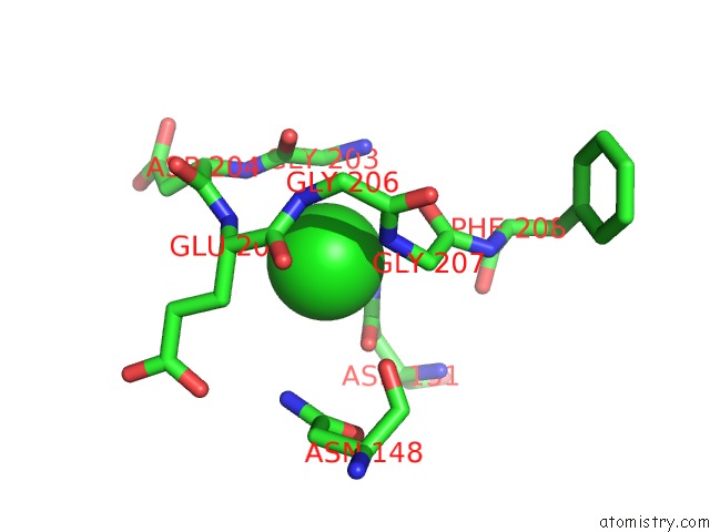
Mono view
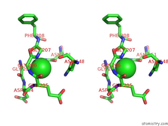
Stereo pair view
Mono view
Stereo pair view
A full contact list of Chlorine with other atoms in the Cl binding
site number 1 of Crystal Structure of Enolase From Lactobacillus Gasseri within 5.0Å range:
|
Reference:
K.Raghunathan,
P.T.Harris,
R.R.Spurbeck,
C.G.Arvidson,
D.N.Arvidson.
Crystal Structure of An Efficacious Gonococcal Adherence Inhibitor: An Enolase From Lactobacillus Gasseri. Febs Lett. V. 588 2212 2014.
ISSN: ISSN 0014-5793
PubMed: 24859038
DOI: 10.1016/J.FEBSLET.2014.05.020
Page generated: Fri Jul 11 19:10:58 2025
ISSN: ISSN 0014-5793
PubMed: 24859038
DOI: 10.1016/J.FEBSLET.2014.05.020
Last articles
Fe in 2YXOFe in 2YRS
Fe in 2YXC
Fe in 2YNM
Fe in 2YVJ
Fe in 2YP1
Fe in 2YU2
Fe in 2YU1
Fe in 2YQB
Fe in 2YOO