Chlorine »
PDB 4msh-4myq »
4mva »
Chlorine in PDB 4mva: 1.43 Angstrom Resolution Crystal Structure of Triosephosphate Isomerase (Tpia) From Escherichia Coli in Complex with Acetyl Phosphate.
Enzymatic activity of 1.43 Angstrom Resolution Crystal Structure of Triosephosphate Isomerase (Tpia) From Escherichia Coli in Complex with Acetyl Phosphate.
All present enzymatic activity of 1.43 Angstrom Resolution Crystal Structure of Triosephosphate Isomerase (Tpia) From Escherichia Coli in Complex with Acetyl Phosphate.:
5.3.1.1;
5.3.1.1;
Protein crystallography data
The structure of 1.43 Angstrom Resolution Crystal Structure of Triosephosphate Isomerase (Tpia) From Escherichia Coli in Complex with Acetyl Phosphate., PDB code: 4mva
was solved by
G.Minasov,
M.L.Kuhn,
I.Dubrovska,
J.Winsor,
L.Shuvalova,
S.Grimshaw,
K.Kwon,
W.F.Anderson,
Center For Structural Genomics Of Infectiousdiseases (Csgid),
with X-Ray Crystallography technique. A brief refinement statistics is given in the table below:
| Resolution Low / High (Å) | 29.24 / 1.43 |
| Space group | P 21 21 21 |
| Cell size a, b, c (Å), α, β, γ (°) | 46.535, 67.529, 150.336, 90.00, 90.00, 90.00 |
| R / Rfree (%) | 13.5 / 15.6 |
Chlorine Binding Sites:
The binding sites of Chlorine atom in the 1.43 Angstrom Resolution Crystal Structure of Triosephosphate Isomerase (Tpia) From Escherichia Coli in Complex with Acetyl Phosphate.
(pdb code 4mva). This binding sites where shown within
5.0 Angstroms radius around Chlorine atom.
In total 2 binding sites of Chlorine where determined in the 1.43 Angstrom Resolution Crystal Structure of Triosephosphate Isomerase (Tpia) From Escherichia Coli in Complex with Acetyl Phosphate., PDB code: 4mva:
Jump to Chlorine binding site number: 1; 2;
In total 2 binding sites of Chlorine where determined in the 1.43 Angstrom Resolution Crystal Structure of Triosephosphate Isomerase (Tpia) From Escherichia Coli in Complex with Acetyl Phosphate., PDB code: 4mva:
Jump to Chlorine binding site number: 1; 2;
Chlorine binding site 1 out of 2 in 4mva
Go back to
Chlorine binding site 1 out
of 2 in the 1.43 Angstrom Resolution Crystal Structure of Triosephosphate Isomerase (Tpia) From Escherichia Coli in Complex with Acetyl Phosphate.
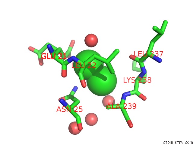
Mono view
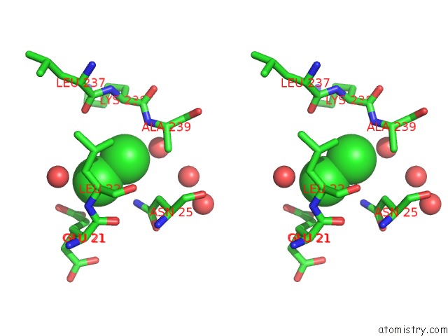
Stereo pair view
Mono view
Stereo pair view
A full contact list of Chlorine with other atoms in the Cl binding
site number 1 of 1.43 Angstrom Resolution Crystal Structure of Triosephosphate Isomerase (Tpia) From Escherichia Coli in Complex with Acetyl Phosphate. within 5.0Å range:
|
Chlorine binding site 2 out of 2 in 4mva
Go back to
Chlorine binding site 2 out
of 2 in the 1.43 Angstrom Resolution Crystal Structure of Triosephosphate Isomerase (Tpia) From Escherichia Coli in Complex with Acetyl Phosphate.
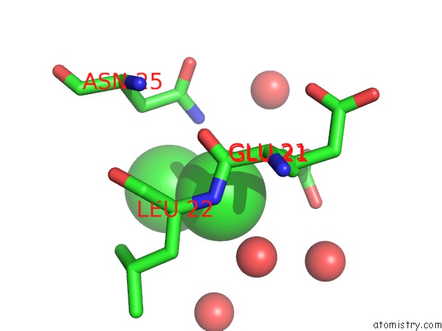
Mono view
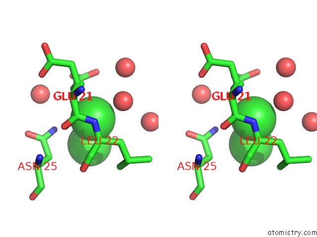
Stereo pair view
Mono view
Stereo pair view
A full contact list of Chlorine with other atoms in the Cl binding
site number 2 of 1.43 Angstrom Resolution Crystal Structure of Triosephosphate Isomerase (Tpia) From Escherichia Coli in Complex with Acetyl Phosphate. within 5.0Å range:
|
Reference:
M.L.Kuhn,
B.Zemaitaitis,
L.I.Hu,
A.Sahu,
D.Sorensen,
G.Minasov,
B.P.Lima,
M.Scholle,
M.Mrksich,
W.F.Anderson,
B.W.Gibson,
B.Schilling,
A.J.Wolfe.
Structural, Kinetic and Proteomic Characterization of Acetyl Phosphate-Dependent Bacterial Protein Acetylation. Plos One V. 9 94816 2014.
ISSN: ESSN 1932-6203
PubMed: 24756028
DOI: 10.1371/JOURNAL.PONE.0094816
Page generated: Fri Jul 11 19:18:34 2025
ISSN: ESSN 1932-6203
PubMed: 24756028
DOI: 10.1371/JOURNAL.PONE.0094816
Last articles
Fe in 2YXOFe in 2YRS
Fe in 2YXC
Fe in 2YNM
Fe in 2YVJ
Fe in 2YP1
Fe in 2YU2
Fe in 2YU1
Fe in 2YQB
Fe in 2YOO