Chlorine »
PDB 4n9a-4ngr »
4n9m »
Chlorine in PDB 4n9m: Joint Neutron/X-Ray Structure of Urate Oxidase in Complex with 8- Hydroxyxanthine
Enzymatic activity of Joint Neutron/X-Ray Structure of Urate Oxidase in Complex with 8- Hydroxyxanthine
All present enzymatic activity of Joint Neutron/X-Ray Structure of Urate Oxidase in Complex with 8- Hydroxyxanthine:
1.7.3.3;
1.7.3.3;
Protein crystallography data
The structure of Joint Neutron/X-Ray Structure of Urate Oxidase in Complex with 8- Hydroxyxanthine, PDB code: 4n9m
was solved by
E.Oksanen,
M.P.Blakeley,
M.Budayova-Spano,
with X-Ray Crystallography technique. A brief refinement statistics is given in the table below:
| Resolution Low / High (Å) | N/A / 2.30 |
| Space group | I 2 2 2 |
| Cell size a, b, c (Å), α, β, γ (°) | 80.180, 96.260, 105.510, 90.00, 90.00, 90.00 |
| R / Rfree (%) | 16.7 / 18.9 |
Other elements in 4n9m:
The structure of Joint Neutron/X-Ray Structure of Urate Oxidase in Complex with 8- Hydroxyxanthine also contains other interesting chemical elements:
| Sodium | (Na) | 1 atom |
Chlorine Binding Sites:
The binding sites of Chlorine atom in the Joint Neutron/X-Ray Structure of Urate Oxidase in Complex with 8- Hydroxyxanthine
(pdb code 4n9m). This binding sites where shown within
5.0 Angstroms radius around Chlorine atom.
In total only one binding site of Chlorine was determined in the Joint Neutron/X-Ray Structure of Urate Oxidase in Complex with 8- Hydroxyxanthine, PDB code: 4n9m:
In total only one binding site of Chlorine was determined in the Joint Neutron/X-Ray Structure of Urate Oxidase in Complex with 8- Hydroxyxanthine, PDB code: 4n9m:
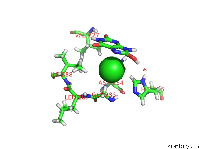
Chlorine binding site 1 out of 1 in 4n9m
Go back to
Chlorine binding site 1 out
of 1 in the Joint Neutron/X-Ray Structure of Urate Oxidase in Complex with 8- Hydroxyxanthine
Mono view

Stereo pair view
Mono view
Stereo pair view
A full contact list of Chlorine with other atoms in the Cl binding
site number 1 of Joint Neutron/X-Ray Structure of Urate Oxidase in Complex with 8- Hydroxyxanthine within 5.0Å range:
|
Reference:
E.Oksanen,
M.P.Blakeley,
M.El-Hajji,
U.Ryde,
M.Budayova-Spano.
The Neutron Structure of Urate Oxidase Resolves A Long-Standing Mechanistic Conundrum and Reveals Unexpected Changes in Protonation. Plos One V. 9 86651 2014.
ISSN: ESSN 1932-6203
PubMed: 24466188
DOI: 10.1371/JOURNAL.PONE.0086651
Page generated: Sun Jul 21 20:33:44 2024
ISSN: ESSN 1932-6203
PubMed: 24466188
DOI: 10.1371/JOURNAL.PONE.0086651
Last articles
Zn in 9J0NZn in 9J0O
Zn in 9J0P
Zn in 9FJX
Zn in 9EKB
Zn in 9C0F
Zn in 9CAH
Zn in 9CH0
Zn in 9CH3
Zn in 9CH1