Chlorine »
PDB 4ngs-4ntd »
4nm3 »
Chlorine in PDB 4nm3: Crystal Structure of Gsk-3/Axin Complex Bound to Phosphorylated N- Terminal Auto-Inhibitory PS9 Peptide
Enzymatic activity of Crystal Structure of Gsk-3/Axin Complex Bound to Phosphorylated N- Terminal Auto-Inhibitory PS9 Peptide
All present enzymatic activity of Crystal Structure of Gsk-3/Axin Complex Bound to Phosphorylated N- Terminal Auto-Inhibitory PS9 Peptide:
2.7.11.1; 2.7.11.26;
2.7.11.1; 2.7.11.26;
Protein crystallography data
The structure of Crystal Structure of Gsk-3/Axin Complex Bound to Phosphorylated N- Terminal Auto-Inhibitory PS9 Peptide, PDB code: 4nm3
was solved by
M.L.-H.Chu,
J.L.Stamos,
M.D.Enos,
N.Shah,
W.I.Weis,
with X-Ray Crystallography technique. A brief refinement statistics is given in the table below:
| Resolution Low / High (Å) | 38.96 / 2.10 |
| Space group | P 61 2 2 |
| Cell size a, b, c (Å), α, β, γ (°) | 81.032, 81.032, 281.085, 90.00, 90.00, 120.00 |
| R / Rfree (%) | 19.4 / 24.2 |
Other elements in 4nm3:
The structure of Crystal Structure of Gsk-3/Axin Complex Bound to Phosphorylated N- Terminal Auto-Inhibitory PS9 Peptide also contains other interesting chemical elements:
| Magnesium | (Mg) | 2 atoms |
Chlorine Binding Sites:
The binding sites of Chlorine atom in the Crystal Structure of Gsk-3/Axin Complex Bound to Phosphorylated N- Terminal Auto-Inhibitory PS9 Peptide
(pdb code 4nm3). This binding sites where shown within
5.0 Angstroms radius around Chlorine atom.
In total only one binding site of Chlorine was determined in the Crystal Structure of Gsk-3/Axin Complex Bound to Phosphorylated N- Terminal Auto-Inhibitory PS9 Peptide, PDB code: 4nm3:
In total only one binding site of Chlorine was determined in the Crystal Structure of Gsk-3/Axin Complex Bound to Phosphorylated N- Terminal Auto-Inhibitory PS9 Peptide, PDB code: 4nm3:
Chlorine binding site 1 out of 1 in 4nm3
Go back to
Chlorine binding site 1 out
of 1 in the Crystal Structure of Gsk-3/Axin Complex Bound to Phosphorylated N- Terminal Auto-Inhibitory PS9 Peptide
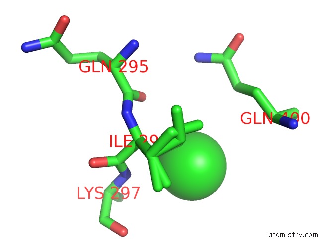
Mono view
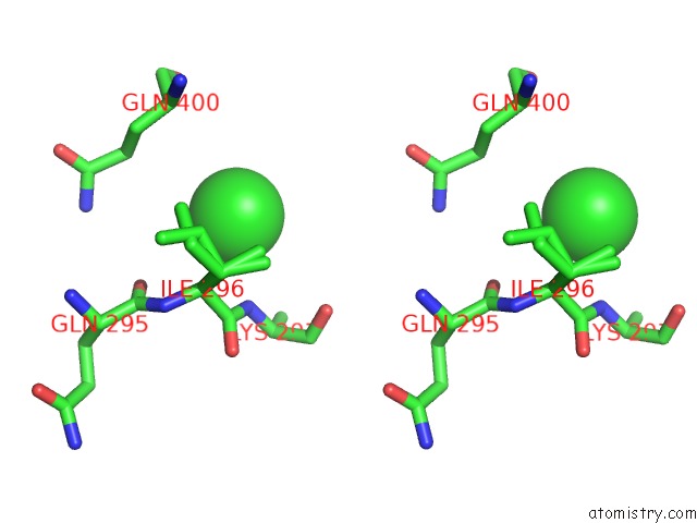
Stereo pair view
Mono view
Stereo pair view
A full contact list of Chlorine with other atoms in the Cl binding
site number 1 of Crystal Structure of Gsk-3/Axin Complex Bound to Phosphorylated N- Terminal Auto-Inhibitory PS9 Peptide within 5.0Å range:
|
Reference:
J.L.Stamos,
M.L.Chu,
M.D.Enos,
N.Shah,
W.I.Weis.
Structural Basis of Gsk-3 Inhibition By N-Terminal Phosphorylation and By the Wnt Receptor LRP6. Elife V. 3 01998 2014.
ISSN: ESSN 2050-084X
PubMed: 24642411
Page generated: Fri Jul 11 19:39:44 2025
ISSN: ESSN 2050-084X
PubMed: 24642411
Last articles
Fe in 2YXOFe in 2YRS
Fe in 2YXC
Fe in 2YNM
Fe in 2YVJ
Fe in 2YP1
Fe in 2YU2
Fe in 2YU1
Fe in 2YQB
Fe in 2YOO