Chlorine »
PDB 4oak-4ogt »
4oej »
Chlorine in PDB 4oej: Structure of Membrane Binding Protein Pleurotolysin B From Pleurotus Ostreatus
Protein crystallography data
The structure of Structure of Membrane Binding Protein Pleurotolysin B From Pleurotus Ostreatus, PDB code: 4oej
was solved by
M.A.Dunstone,
T.T.Caradoc-Davies,
J.C.Whisstock,
R.H.P.Law,
with X-Ray Crystallography technique. A brief refinement statistics is given in the table below:
| Resolution Low / High (Å) | 26.56 / 2.20 |
| Space group | P 31 2 1 |
| Cell size a, b, c (Å), α, β, γ (°) | 71.580, 71.580, 174.920, 90.00, 90.00, 120.00 |
| R / Rfree (%) | 18.9 / 22.1 |
Chlorine Binding Sites:
The binding sites of Chlorine atom in the Structure of Membrane Binding Protein Pleurotolysin B From Pleurotus Ostreatus
(pdb code 4oej). This binding sites where shown within
5.0 Angstroms radius around Chlorine atom.
In total only one binding site of Chlorine was determined in the Structure of Membrane Binding Protein Pleurotolysin B From Pleurotus Ostreatus, PDB code: 4oej:
In total only one binding site of Chlorine was determined in the Structure of Membrane Binding Protein Pleurotolysin B From Pleurotus Ostreatus, PDB code: 4oej:
Chlorine binding site 1 out of 1 in 4oej
Go back to
Chlorine binding site 1 out
of 1 in the Structure of Membrane Binding Protein Pleurotolysin B From Pleurotus Ostreatus
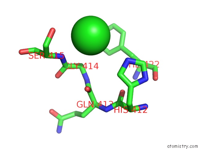
Mono view
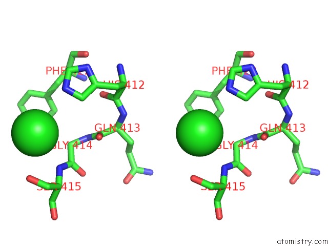
Stereo pair view
Mono view
Stereo pair view
A full contact list of Chlorine with other atoms in the Cl binding
site number 1 of Structure of Membrane Binding Protein Pleurotolysin B From Pleurotus Ostreatus within 5.0Å range:
|
Reference:
N.Lukoyanova,
S.C.Kondos,
I.Farabella,
R.H.Law,
C.F.Reboul,
T.T.Caradoc-Davies,
B.A.Spicer,
O.Kleifeld,
D.A.Traore,
S.M.Ekkel,
I.Voskoboinik,
J.A.Trapani,
T.Hatfaludi,
K.Oliver,
E.M.Hotze,
R.K.Tweten,
J.C.Whisstock,
M.Topf,
H.R.Saibil,
M.A.Dunstone.
Conformational Changes During Pore Formation By the Perforin-Related Protein Pleurotolysin. Plos Biol. V. 13 02049 2015.
ISSN: ISSN 1544-9173
PubMed: 25654333
DOI: 10.1371/JOURNAL.PBIO.1002049
Page generated: Fri Jul 11 20:01:25 2025
ISSN: ISSN 1544-9173
PubMed: 25654333
DOI: 10.1371/JOURNAL.PBIO.1002049
Last articles
Fe in 2YXOFe in 2YRS
Fe in 2YXC
Fe in 2YNM
Fe in 2YVJ
Fe in 2YP1
Fe in 2YU2
Fe in 2YU1
Fe in 2YQB
Fe in 2YOO