Chlorine »
PDB 4otk-4oz2 »
4oy3 »
Chlorine in PDB 4oy3: Crystal Structure of the Helicobacter Pylori Mtan-D198N Mutant with S- Adenosylhomocysteine in the Active Site
Enzymatic activity of Crystal Structure of the Helicobacter Pylori Mtan-D198N Mutant with S- Adenosylhomocysteine in the Active Site
All present enzymatic activity of Crystal Structure of the Helicobacter Pylori Mtan-D198N Mutant with S- Adenosylhomocysteine in the Active Site:
3.2.2.30; 3.2.2.9;
3.2.2.30; 3.2.2.9;
Protein crystallography data
The structure of Crystal Structure of the Helicobacter Pylori Mtan-D198N Mutant with S- Adenosylhomocysteine in the Active Site, PDB code: 4oy3
was solved by
V.Mishra,
D.R.Ronning,
with X-Ray Crystallography technique. A brief refinement statistics is given in the table below:
| Resolution Low / High (Å) | 22.02 / 1.20 |
| Space group | P 32 2 1 |
| Cell size a, b, c (Å), α, β, γ (°) | 80.733, 80.733, 67.199, 90.00, 90.00, 120.00 |
| R / Rfree (%) | 13 / 14.5 |
Chlorine Binding Sites:
The binding sites of Chlorine atom in the Crystal Structure of the Helicobacter Pylori Mtan-D198N Mutant with S- Adenosylhomocysteine in the Active Site
(pdb code 4oy3). This binding sites where shown within
5.0 Angstroms radius around Chlorine atom.
In total only one binding site of Chlorine was determined in the Crystal Structure of the Helicobacter Pylori Mtan-D198N Mutant with S- Adenosylhomocysteine in the Active Site, PDB code: 4oy3:
In total only one binding site of Chlorine was determined in the Crystal Structure of the Helicobacter Pylori Mtan-D198N Mutant with S- Adenosylhomocysteine in the Active Site, PDB code: 4oy3:
Chlorine binding site 1 out of 1 in 4oy3
Go back to
Chlorine binding site 1 out
of 1 in the Crystal Structure of the Helicobacter Pylori Mtan-D198N Mutant with S- Adenosylhomocysteine in the Active Site
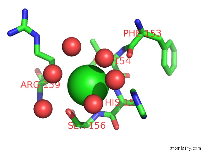
Mono view
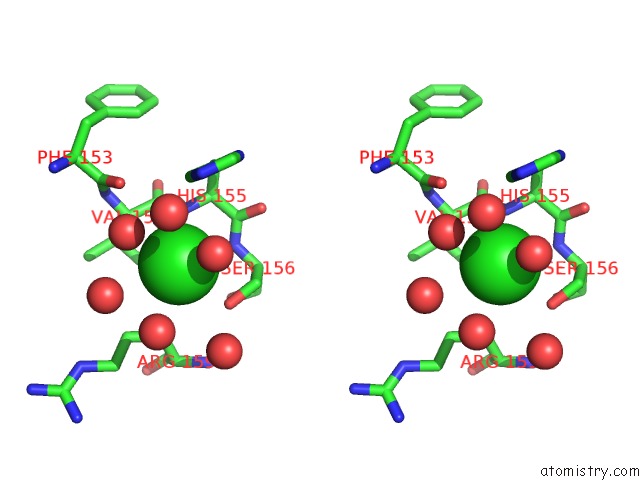
Stereo pair view
Mono view
Stereo pair view
A full contact list of Chlorine with other atoms in the Cl binding
site number 1 of Crystal Structure of the Helicobacter Pylori Mtan-D198N Mutant with S- Adenosylhomocysteine in the Active Site within 5.0Å range:
|
Reference:
V.Mishra,
D.R.Ronning.
Crystal Structures of the Helicobacter Pylori Mtan Enzyme Reveal Specific Interactions Between S-Adenosylhomocysteine and the 5'-Alkylthio Binding Subsite. Biochemistry V. 51 9763 2012.
ISSN: ISSN 0006-2960
PubMed: 23148563
DOI: 10.1021/BI301221K
Page generated: Thu Jul 25 23:40:59 2024
ISSN: ISSN 0006-2960
PubMed: 23148563
DOI: 10.1021/BI301221K
Last articles
Zn in 9J0NZn in 9J0O
Zn in 9J0P
Zn in 9FJX
Zn in 9EKB
Zn in 9C0F
Zn in 9CAH
Zn in 9CH0
Zn in 9CH3
Zn in 9CH1