Chlorine »
PDB 4otk-4oz2 »
4oyw »
Chlorine in PDB 4oyw: Crystal Structure of Human Soluble Adenylate Cyclase
Enzymatic activity of Crystal Structure of Human Soluble Adenylate Cyclase
All present enzymatic activity of Crystal Structure of Human Soluble Adenylate Cyclase:
4.6.1.1;
4.6.1.1;
Protein crystallography data
The structure of Crystal Structure of Human Soluble Adenylate Cyclase, PDB code: 4oyw
was solved by
M.Vinkovic,
with X-Ray Crystallography technique. A brief refinement statistics is given in the table below:
| Resolution Low / High (Å) | 49.71 / 1.70 |
| Space group | P 63 |
| Cell size a, b, c (Å), α, β, γ (°) | 99.424, 99.424, 97.391, 90.00, 90.00, 120.00 |
| R / Rfree (%) | 18.2 / 21.1 |
Chlorine Binding Sites:
The binding sites of Chlorine atom in the Crystal Structure of Human Soluble Adenylate Cyclase
(pdb code 4oyw). This binding sites where shown within
5.0 Angstroms radius around Chlorine atom.
In total only one binding site of Chlorine was determined in the Crystal Structure of Human Soluble Adenylate Cyclase, PDB code: 4oyw:
In total only one binding site of Chlorine was determined in the Crystal Structure of Human Soluble Adenylate Cyclase, PDB code: 4oyw:
Chlorine binding site 1 out of 1 in 4oyw
Go back to
Chlorine binding site 1 out
of 1 in the Crystal Structure of Human Soluble Adenylate Cyclase
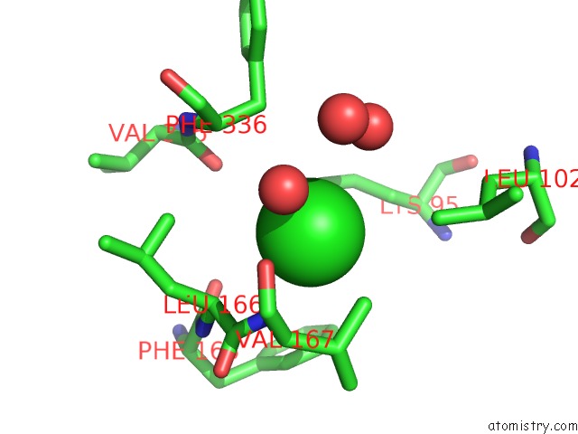
Mono view
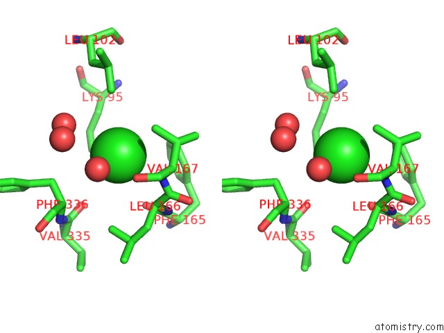
Stereo pair view
Mono view
Stereo pair view
A full contact list of Chlorine with other atoms in the Cl binding
site number 1 of Crystal Structure of Human Soluble Adenylate Cyclase within 5.0Å range:
|
Reference:
S.M.Saalau-Bethell,
V.Berdini,
A.Cleasby,
M.Congreve,
J.E.Coyle,
V.Lock,
C.W.Murray,
M.A.O'brien,
S.J.Rich,
T.Sambrook,
M.Vinkovic,
J.R.Yon,
H.Jhoti.
Crystal Structure of Human Soluble Adenylate Cyclase Reveals A Distinct, Highly Flexible Allosteric Bicarbonate Binding Pocket. Chemmedchem V. 9 823 2014.
ISSN: ESSN 1860-7187
PubMed: 24616449
DOI: 10.1002/CMDC.201300480
Page generated: Thu Jul 25 23:43:03 2024
ISSN: ESSN 1860-7187
PubMed: 24616449
DOI: 10.1002/CMDC.201300480
Last articles
Zn in 9J0NZn in 9J0O
Zn in 9J0P
Zn in 9FJX
Zn in 9EKB
Zn in 9C0F
Zn in 9CAH
Zn in 9CH0
Zn in 9CH3
Zn in 9CH1