Chlorine in PDB 4p18: Crystal Structure of Frog M Ferritin Mutant D80K
Enzymatic activity of Crystal Structure of Frog M Ferritin Mutant D80K
All present enzymatic activity of Crystal Structure of Frog M Ferritin Mutant D80K:
1.16.3.1;
Protein crystallography data
The structure of Crystal Structure of Frog M Ferritin Mutant D80K, PDB code: 4p18
was solved by
C.Pozzi,
F.Di Pisa,
S.Mangani,
C.Bernacchioni,
V.Ghini,
P.Turano,
with X-Ray Crystallography technique. A brief refinement statistics is given in the table below:
Resolution Low / High (Å)
|
75.35 /
1.91
|
Space group
|
C 1 2 1
|
Cell size a, b, c (Å), α, β, γ (°)
|
238.930,
238.430,
119.690,
90.00,
94.33,
90.00
|
R / Rfree (%)
|
19.6 /
23.1
|
Chlorine Binding Sites:
Chlorine binding site 1 out
of 31 in 4p18
Go back to
Chlorine Binding Sites List in 4p18
Chlorine binding site 1 out
of 31 in the Crystal Structure of Frog M Ferritin Mutant D80K
Mono view
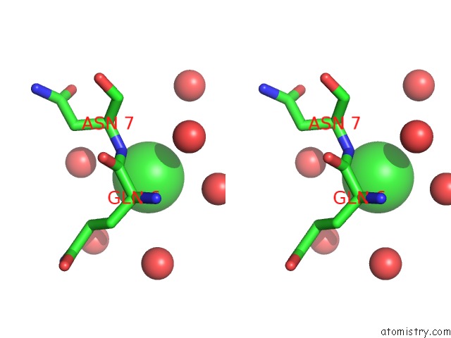
Stereo pair view
|
A full contact list of Chlorine with other atoms in the Cl binding
site number 1 of Crystal Structure of Frog M Ferritin Mutant D80K within 5.0Å range:
probe
|
atom
|
residue
|
distance (Å)
|
B
|
Occ
|
B:Cl201
b:29.7
occ:1.00
|
O
|
R:HOH406
|
3.4
|
15.2
|
1.0
|
N
|
B:ASN7
|
3.4
|
5.2
|
1.0
|
O
|
B:HOH334
|
3.8
|
9.4
|
1.0
|
O
|
B:HOH438
|
3.8
|
29.0
|
1.0
|
CA
|
B:GLN6
|
4.2
|
5.0
|
1.0
|
CA
|
B:ASN7
|
4.2
|
5.4
|
1.0
|
O
|
R:HOH396
|
4.2
|
9.7
|
1.0
|
C
|
B:GLN6
|
4.3
|
5.1
|
1.0
|
O
|
B:HOH375
|
4.5
|
16.2
|
1.0
|
CG
|
B:GLN6
|
4.5
|
5.0
|
1.0
|
O
|
B:HOH429
|
4.9
|
26.2
|
1.0
|
CB
|
B:GLN6
|
4.9
|
4.9
|
1.0
|
|
Chlorine binding site 2 out
of 31 in 4p18
Go back to
Chlorine Binding Sites List in 4p18
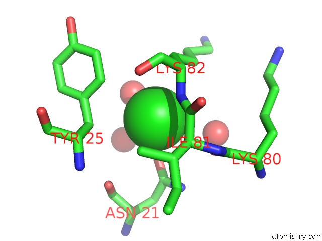
Chlorine binding site 2 out
of 31 in the Crystal Structure of Frog M Ferritin Mutant D80K
Mono view
Stereo pair view
|
A full contact list of Chlorine with other atoms in the Cl binding
site number 2 of Crystal Structure of Frog M Ferritin Mutant D80K within 5.0Å range:
probe
|
atom
|
residue
|
distance (Å)
|
B
|
Occ
|
B:Cl202
b:23.7
occ:1.00
|
OD1
|
B:ASN21
|
2.8
|
4.4
|
1.0
|
N
|
B:LYS82
|
2.9
|
7.0
|
1.0
|
O
|
B:HOH446
|
3.0
|
22.5
|
1.0
|
CB
|
B:LYS82
|
3.6
|
8.4
|
1.0
|
O
|
B:LYS80
|
3.6
|
6.2
|
1.0
|
CB
|
B:TYR25
|
3.6
|
4.1
|
1.0
|
CA
|
B:ILE81
|
3.7
|
6.2
|
1.0
|
CG
|
B:LYS82
|
3.7
|
9.7
|
1.0
|
CD1
|
B:TYR25
|
3.7
|
4.3
|
1.0
|
C
|
B:ILE81
|
3.8
|
6.5
|
1.0
|
CG
|
B:ASN21
|
3.8
|
4.3
|
1.0
|
CA
|
B:LYS82
|
3.8
|
7.7
|
1.0
|
O
|
B:HOH368
|
3.8
|
10.4
|
1.0
|
O
|
B:HOH500
|
4.0
|
11.5
|
1.0
|
ND2
|
B:ASN21
|
4.0
|
4.3
|
1.0
|
CG
|
B:TYR25
|
4.1
|
4.2
|
1.0
|
CD
|
B:LYS82
|
4.3
|
10.8
|
1.0
|
CG2
|
B:ILE81
|
4.4
|
5.8
|
1.0
|
O
|
B:ASN21
|
4.5
|
4.0
|
1.0
|
C
|
B:LYS80
|
4.5
|
6.5
|
1.0
|
O
|
B:LYS82
|
4.5
|
7.2
|
1.0
|
N
|
B:ILE81
|
4.6
|
6.2
|
1.0
|
CA
|
B:TYR25
|
4.6
|
3.9
|
1.0
|
CB
|
B:ILE81
|
4.6
|
6.0
|
1.0
|
C
|
B:LYS82
|
4.7
|
7.6
|
1.0
|
CE1
|
B:TYR25
|
4.8
|
4.4
|
1.0
|
O
|
B:ILE81
|
5.0
|
6.2
|
1.0
|
N
|
B:TYR25
|
5.0
|
3.9
|
1.0
|
|
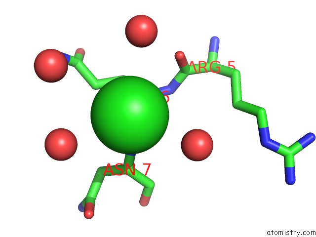
Chlorine binding site 3 out
of 31 in 4p18
Go back to
Chlorine Binding Sites List in 4p18
Chlorine binding site 3 out
of 31 in the Crystal Structure of Frog M Ferritin Mutant D80K
Mono view
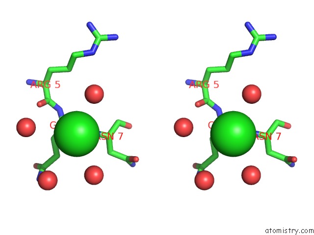
Stereo pair view
|
A full contact list of Chlorine with other atoms in the Cl binding
site number 3 of Crystal Structure of Frog M Ferritin Mutant D80K within 5.0Å range:
probe
|
atom
|
residue
|
distance (Å)
|
B
|
Occ
|
C:Cl202
b:32.0
occ:1.00
|
N
|
C:ASN7
|
3.4
|
6.0
|
1.0
|
O
|
C:HOH330
|
3.8
|
7.5
|
1.0
|
O
|
F:HOH338
|
3.8
|
12.0
|
1.0
|
CA
|
C:GLN6
|
4.1
|
6.0
|
1.0
|
CA
|
C:ASN7
|
4.2
|
6.1
|
1.0
|
O
|
F:HOH378
|
4.2
|
10.2
|
1.0
|
O
|
C:HOH402
|
4.2
|
23.5
|
1.0
|
C
|
C:GLN6
|
4.3
|
6.0
|
1.0
|
CG
|
C:GLN6
|
4.5
|
6.0
|
1.0
|
O
|
C:ARG5
|
4.8
|
6.5
|
1.0
|
CB
|
C:GLN6
|
4.9
|
6.0
|
1.0
|
|
Chlorine binding site 4 out
of 31 in 4p18
Go back to
Chlorine Binding Sites List in 4p18
Chlorine binding site 4 out
of 31 in the Crystal Structure of Frog M Ferritin Mutant D80K
Mono view
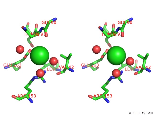
Stereo pair view
|
A full contact list of Chlorine with other atoms in the Cl binding
site number 4 of Crystal Structure of Frog M Ferritin Mutant D80K within 5.0Å range:
probe
|
atom
|
residue
|
distance (Å)
|
B
|
Occ
|
D:Cl203
b:36.9
occ:1.00
|
OE1
|
D:GLU163
|
2.9
|
8.0
|
1.0
|
O
|
D:HOH395
|
2.9
|
22.4
|
1.0
|
OE2
|
D:GLU163
|
3.3
|
7.2
|
1.0
|
NH2
|
F:ARG153
|
3.4
|
5.6
|
1.0
|
CD
|
D:GLU163
|
3.4
|
7.8
|
1.0
|
CB
|
D:ASN91
|
3.8
|
6.8
|
1.0
|
O
|
D:HOH441
|
3.8
|
24.8
|
1.0
|
O
|
F:HOH387
|
4.0
|
23.7
|
1.0
|
O
|
D:GLY90
|
4.0
|
7.1
|
1.0
|
CA
|
D:ASN91
|
4.1
|
6.5
|
1.0
|
CD1
|
D:LEU44
|
4.3
|
5.2
|
1.0
|
CZ
|
F:ARG153
|
4.4
|
5.7
|
1.0
|
NE
|
F:ARG153
|
4.5
|
5.8
|
1.0
|
CG1
|
D:VAL42
|
4.6
|
5.7
|
1.0
|
CG
|
D:GLU163
|
4.7
|
7.6
|
1.0
|
C
|
D:GLY90
|
4.9
|
7.1
|
1.0
|
N
|
D:THR92
|
5.0
|
6.1
|
1.0
|
N
|
D:ASN91
|
5.0
|
6.8
|
1.0
|
|
Chlorine binding site 5 out
of 31 in 4p18
Go back to
Chlorine Binding Sites List in 4p18
Chlorine binding site 5 out
of 31 in the Crystal Structure of Frog M Ferritin Mutant D80K
Mono view
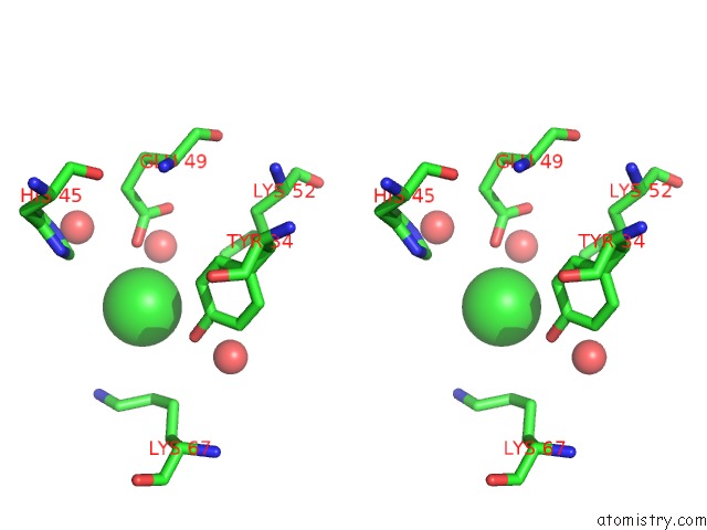
Stereo pair view
|
A full contact list of Chlorine with other atoms in the Cl binding
site number 5 of Crystal Structure of Frog M Ferritin Mutant D80K within 5.0Å range:
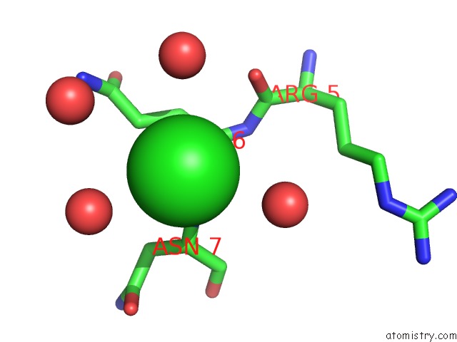
probe
|
atom
|
residue
|
distance (Å)
|
B
|
Occ
|
D:Cl204
b:38.6
occ:1.00
|
OE2
|
D:GLU49
|
3.1
|
12.7
|
1.0
|
O
|
D:HOH433
|
3.6
|
30.4
|
1.0
|
OH
|
D:TYR34
|
3.7
|
7.5
|
0.7
|
CD
|
C:LYS67
|
4.1
|
11.5
|
0.7
|
CD
|
D:GLU49
|
4.2
|
11.4
|
1.0
|
NZ
|
C:LYS67
|
4.3
|
12.7
|
0.7
|
CE
|
D:LYS52
|
4.3
|
11.3
|
1.0
|
NZ
|
D:LYS52
|
4.4
|
12.6
|
1.0
|
CE1
|
D:TYR34
|
4.4
|
6.8
|
0.7
|
O
|
D:HOH488
|
4.5
|
39.5
|
1.0
|
CZ
|
D:TYR34
|
4.5
|
7.1
|
0.7
|
OE1
|
D:GLU49
|
4.5
|
12.4
|
1.0
|
CE
|
C:LYS67
|
4.5
|
11.6
|
0.7
|
CE1
|
D:HIS45
|
4.5
|
8.5
|
1.0
|
O
|
D:HOH405
|
4.6
|
23.8
|
1.0
|
CG
|
C:LYS67
|
4.8
|
10.9
|
1.0
|
|
Chlorine binding site 6 out
of 31 in 4p18
Go back to
Chlorine Binding Sites List in 4p18
Chlorine binding site 6 out
of 31 in the Crystal Structure of Frog M Ferritin Mutant D80K
Mono view
Stereo pair view
|
A full contact list of Chlorine with other atoms in the Cl binding
site number 6 of Crystal Structure of Frog M Ferritin Mutant D80K within 5.0Å range:
probe
|
atom
|
residue
|
distance (Å)
|
B
|
Occ
|
M:Cl201
b:32.7
occ:1.00
|
N
|
M:ASN7
|
3.4
|
6.7
|
1.0
|
O
|
D:HOH336
|
3.7
|
12.5
|
1.0
|
O
|
M:HOH336
|
3.8
|
7.0
|
1.0
|
O
|
M:HOH352
|
3.9
|
13.9
|
1.0
|
CA
|
M:GLN6
|
4.1
|
6.6
|
1.0
|
CA
|
M:ASN7
|
4.2
|
6.8
|
1.0
|
O
|
D:HOH353
|
4.2
|
12.6
|
1.0
|
C
|
M:GLN6
|
4.3
|
6.7
|
1.0
|
CG
|
M:GLN6
|
4.5
|
6.7
|
1.0
|
CB
|
M:GLN6
|
4.9
|
6.6
|
1.0
|
O
|
M:ARG5
|
5.0
|
7.0
|
1.0
|
|
Chlorine binding site 7 out
of 31 in 4p18
Go back to
Chlorine Binding Sites List in 4p18
Chlorine binding site 7 out
of 31 in the Crystal Structure of Frog M Ferritin Mutant D80K
Mono view
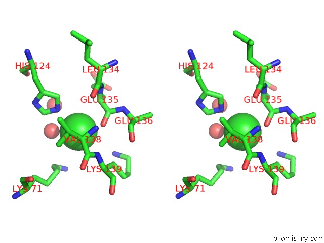
Stereo pair view
|
A full contact list of Chlorine with other atoms in the Cl binding
site number 7 of Crystal Structure of Frog M Ferritin Mutant D80K within 5.0Å range:
probe
|
atom
|
residue
|
distance (Å)
|
B
|
Occ
|
O:Cl202
b:36.6
occ:1.00
|
O
|
O:GLU135
|
2.8
|
9.2
|
1.0
|
NE2
|
W:HIS124
|
3.0
|
4.8
|
1.0
|
O
|
W:HOH451
|
3.2
|
23.1
|
1.0
|
C
|
O:GLU135
|
3.5
|
9.1
|
1.0
|
CA
|
O:GLU135
|
3.6
|
9.4
|
1.0
|
CE1
|
W:HIS124
|
3.7
|
4.8
|
1.0
|
CG1
|
O:VAL138
|
3.8
|
5.9
|
1.0
|
CB
|
O:VAL138
|
3.8
|
6.2
|
1.0
|
N
|
O:LYS139
|
4.0
|
6.0
|
1.0
|
CB
|
O:GLU135
|
4.0
|
10.7
|
1.0
|
CD
|
W:LYS71
|
4.2
|
7.3
|
1.0
|
CD2
|
W:HIS124
|
4.2
|
4.8
|
1.0
|
CE
|
W:LYS71
|
4.3
|
7.5
|
1.0
|
NZ
|
O:LYS139
|
4.4
|
8.8
|
0.7
|
O
|
W:HOH511
|
4.4
|
22.5
|
1.0
|
NZ
|
W:LYS71
|
4.5
|
7.6
|
1.0
|
CB
|
O:LYS139
|
4.5
|
6.8
|
1.0
|
C
|
O:VAL138
|
4.6
|
6.3
|
1.0
|
CA
|
O:VAL138
|
4.7
|
6.4
|
1.0
|
CA
|
O:LYS139
|
4.7
|
6.3
|
1.0
|
CG
|
O:GLU135
|
4.7
|
12.1
|
1.0
|
N
|
O:GLU136
|
4.8
|
8.6
|
1.0
|
O
|
O:LEU134
|
4.9
|
8.2
|
1.0
|
N
|
O:GLU135
|
4.9
|
8.9
|
1.0
|
CG2
|
O:VAL138
|
4.9
|
6.2
|
1.0
|
ND1
|
W:HIS124
|
5.0
|
4.8
|
1.0
|
N
|
O:VAL138
|
5.0
|
6.9
|
1.0
|
|
Chlorine binding site 8 out
of 31 in 4p18
Go back to
Chlorine Binding Sites List in 4p18
Chlorine binding site 8 out
of 31 in the Crystal Structure of Frog M Ferritin Mutant D80K
Mono view
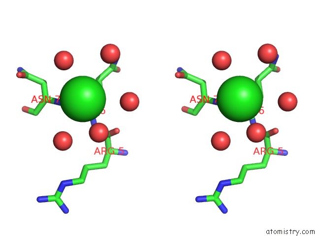
Stereo pair view
|
A full contact list of Chlorine with other atoms in the Cl binding
site number 8 of Crystal Structure of Frog M Ferritin Mutant D80K within 5.0Å range:
probe
|
atom
|
residue
|
distance (Å)
|
B
|
Occ
|
O:Cl203
b:38.0
occ:1.00
|
O
|
O:HOH482
|
2.9
|
23.9
|
1.0
|
N
|
O:ASN7
|
3.5
|
6.2
|
1.0
|
O
|
N:HOH394
|
3.7
|
14.4
|
1.0
|
O
|
O:HOH354
|
3.9
|
11.2
|
1.0
|
O
|
O:HOH389
|
4.0
|
17.1
|
1.0
|
O
|
N:HOH356
|
4.1
|
11.3
|
1.0
|
CA
|
O:GLN6
|
4.2
|
6.6
|
1.0
|
C
|
O:GLN6
|
4.4
|
6.5
|
1.0
|
CA
|
O:ASN7
|
4.4
|
6.3
|
1.0
|
CG
|
O:GLN6
|
4.5
|
6.8
|
1.0
|
CB
|
O:GLN6
|
4.9
|
6.7
|
1.0
|
O
|
O:ARG5
|
4.9
|
7.5
|
1.0
|
|
Chlorine binding site 9 out
of 31 in 4p18
Go back to
Chlorine Binding Sites List in 4p18
Chlorine binding site 9 out
of 31 in the Crystal Structure of Frog M Ferritin Mutant D80K
Mono view
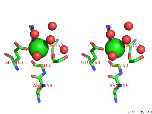
Stereo pair view
|
A full contact list of Chlorine with other atoms in the Cl binding
site number 9 of Crystal Structure of Frog M Ferritin Mutant D80K within 5.0Å range:
probe
|
atom
|
residue
|
distance (Å)
|
B
|
Occ
|
S:Cl201
b:25.2
occ:1.00
|
OE1
|
G:GLU163
|
2.6
|
4.7
|
1.0
|
O2
|
G:EDO205
|
2.8
|
7.3
|
1.0
|
NE
|
S:ARG153
|
2.9
|
4.0
|
1.0
|
O
|
G:HOH336
|
3.2
|
10.5
|
1.0
|
O
|
G:HOH433
|
3.2
|
24.5
|
1.0
|
CG
|
S:ARG153
|
3.4
|
4.3
|
1.0
|
CA
|
G:GLY160
|
3.5
|
3.6
|
1.0
|
CD
|
S:ARG153
|
3.5
|
4.1
|
1.0
|
CD
|
G:GLU163
|
3.6
|
4.5
|
1.0
|
CB
|
G:GLU163
|
3.7
|
4.1
|
1.0
|
C2
|
G:EDO205
|
3.8
|
7.3
|
1.0
|
CZ
|
S:ARG153
|
3.9
|
4.0
|
1.0
|
NH2
|
S:ARG153
|
4.0
|
4.0
|
1.0
|
CG
|
G:GLU163
|
4.0
|
4.3
|
1.0
|
O
|
G:GLY160
|
4.1
|
3.5
|
1.0
|
CB
|
S:ARG153
|
4.1
|
4.1
|
1.0
|
O
|
G:HOH363
|
4.1
|
9.1
|
1.0
|
C
|
G:GLY160
|
4.3
|
3.5
|
1.0
|
O
|
S:HOH351
|
4.5
|
18.7
|
1.0
|
N
|
G:GLY160
|
4.6
|
3.6
|
1.0
|
OE2
|
G:GLU163
|
4.6
|
4.5
|
1.0
|
O
|
G:HOH399
|
4.6
|
12.3
|
1.0
|
CA
|
S:ARG153
|
4.8
|
4.0
|
1.0
|
C1
|
G:EDO205
|
4.9
|
7.5
|
1.0
|
O
|
G:ASN159
|
5.0
|
3.6
|
1.0
|
|
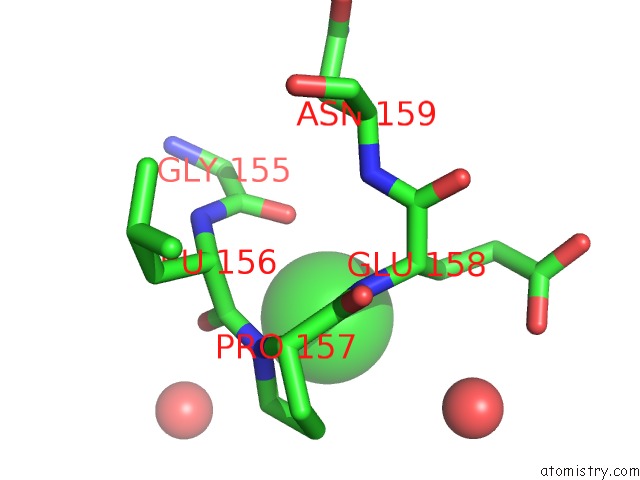
Chlorine binding site 10 out
of 31 in 4p18
Go back to
Chlorine Binding Sites List in 4p18
Chlorine binding site 10 out
of 31 in the Crystal Structure of Frog M Ferritin Mutant D80K
Mono view
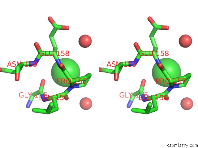
Stereo pair view
|
A full contact list of Chlorine with other atoms in the Cl binding
site number 10 of Crystal Structure of Frog M Ferritin Mutant D80K within 5.0Å range:
probe
|
atom
|
residue
|
distance (Å)
|
B
|
Occ
|
T:Cl203
b:23.2
occ:1.00
|
O
|
T:GLY155
|
3.2
|
5.0
|
1.0
|
O
|
T:LEU156
|
3.3
|
6.2
|
1.0
|
N
|
T:GLU158
|
3.3
|
8.1
|
1.0
|
N
|
T:PRO157
|
3.4
|
6.9
|
1.0
|
C
|
T:LEU156
|
3.5
|
6.2
|
1.0
|
CD
|
T:PRO157
|
3.5
|
7.1
|
1.0
|
CB
|
T:GLU158
|
3.5
|
9.2
|
1.0
|
C
|
T:GLY155
|
4.0
|
4.9
|
1.0
|
CA
|
T:GLU158
|
4.0
|
8.4
|
1.0
|
CG
|
T:PRO157
|
4.2
|
7.1
|
1.0
|
C
|
T:PRO157
|
4.3
|
7.9
|
1.0
|
CA
|
T:PRO157
|
4.4
|
7.3
|
1.0
|
O
|
T:HOH303
|
4.5
|
12.5
|
1.0
|
CA
|
T:LEU156
|
4.5
|
5.7
|
1.0
|
N
|
T:LEU156
|
4.6
|
5.2
|
1.0
|
O
|
T:HOH302
|
4.7
|
11.8
|
1.0
|
CA
|
T:GLY155
|
4.8
|
4.7
|
1.0
|
CG
|
T:GLU158
|
4.9
|
10.1
|
1.0
|
N
|
T:ASN159
|
4.9
|
7.3
|
1.0
|
|
Reference:
C.Bernacchioni,
V.Ghini,
C.Pozzi,
F.Di Pisa,
E.C.Theil,
P.Turano.
Loop Electrostatics Modulates the Intersubunit Interactions in Ferritin. Acs Chem.Biol. 2014.
ISSN: ESSN 1554-8937
PubMed: 25148224
DOI: 10.1021/CB500431R
Page generated: Thu Jul 25 23:46:17 2024
|