Chlorine »
PDB 4pk6-4pws »
4pm4 »
Chlorine in PDB 4pm4: Structure of A Putative Periplasmic Iron Siderophore Binding Protein (RV0265C) From Mycobacterium Tuberculosis H37RV
Protein crystallography data
The structure of Structure of A Putative Periplasmic Iron Siderophore Binding Protein (RV0265C) From Mycobacterium Tuberculosis H37RV, PDB code: 4pm4
was solved by
M.A.Arbing,
S.Chan,
N.Tran,
E.Kuo,
J.Lu,
L.R.Harris,
T.T.Zhou,
D.Eisenberg,
Tb Structural Genomics Consortium (Tbsgc),
with X-Ray Crystallography technique. A brief refinement statistics is given in the table below:
| Resolution Low / High (Å) | 29.20 / 2.20 |
| Space group | P 1 21 1 |
| Cell size a, b, c (Å), α, β, γ (°) | 69.551, 65.434, 72.330, 90.00, 116.41, 90.00 |
| R / Rfree (%) | 20.3 / 25.2 |
Chlorine Binding Sites:
The binding sites of Chlorine atom in the Structure of A Putative Periplasmic Iron Siderophore Binding Protein (RV0265C) From Mycobacterium Tuberculosis H37RV
(pdb code 4pm4). This binding sites where shown within
5.0 Angstroms radius around Chlorine atom.
In total 3 binding sites of Chlorine where determined in the Structure of A Putative Periplasmic Iron Siderophore Binding Protein (RV0265C) From Mycobacterium Tuberculosis H37RV, PDB code: 4pm4:
Jump to Chlorine binding site number: 1; 2; 3;
In total 3 binding sites of Chlorine where determined in the Structure of A Putative Periplasmic Iron Siderophore Binding Protein (RV0265C) From Mycobacterium Tuberculosis H37RV, PDB code: 4pm4:
Jump to Chlorine binding site number: 1; 2; 3;
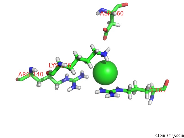
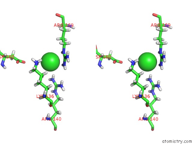
Chlorine binding site 1 out of 3 in 4pm4
Go back to
Chlorine binding site 1 out
of 3 in the Structure of A Putative Periplasmic Iron Siderophore Binding Protein (RV0265C) From Mycobacterium Tuberculosis H37RV
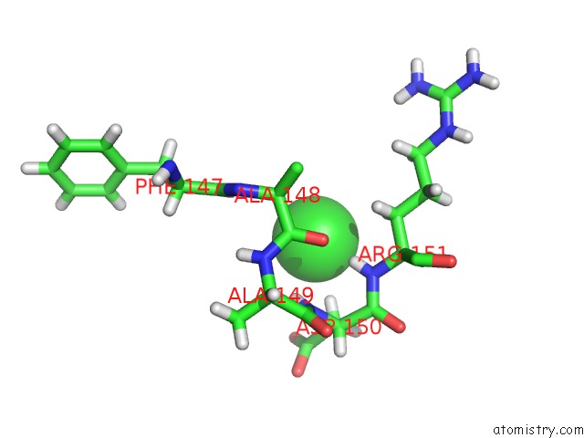
Mono view
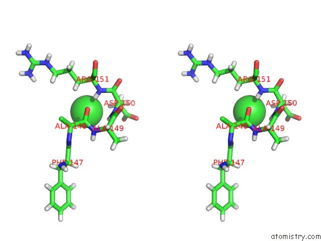
Stereo pair view
Mono view
Stereo pair view
A full contact list of Chlorine with other atoms in the Cl binding
site number 1 of Structure of A Putative Periplasmic Iron Siderophore Binding Protein (RV0265C) From Mycobacterium Tuberculosis H37RV within 5.0Å range:
|
Chlorine binding site 2 out of 3 in 4pm4
Go back to
Chlorine binding site 2 out
of 3 in the Structure of A Putative Periplasmic Iron Siderophore Binding Protein (RV0265C) From Mycobacterium Tuberculosis H37RV
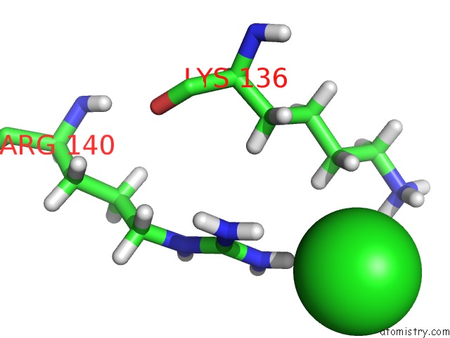
Mono view
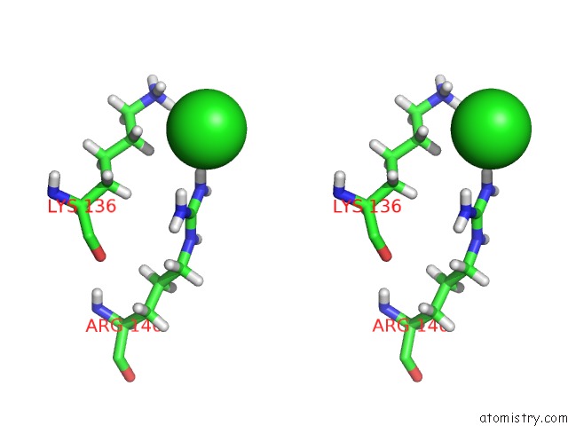
Stereo pair view
Mono view
Stereo pair view
A full contact list of Chlorine with other atoms in the Cl binding
site number 2 of Structure of A Putative Periplasmic Iron Siderophore Binding Protein (RV0265C) From Mycobacterium Tuberculosis H37RV within 5.0Å range:
|
Chlorine binding site 3 out of 3 in 4pm4
Go back to
Chlorine binding site 3 out
of 3 in the Structure of A Putative Periplasmic Iron Siderophore Binding Protein (RV0265C) From Mycobacterium Tuberculosis H37RV
Mono view
Stereo pair view
Mono view
Stereo pair view
A full contact list of Chlorine with other atoms in the Cl binding
site number 3 of Structure of A Putative Periplasmic Iron Siderophore Binding Protein (RV0265C) From Mycobacterium Tuberculosis H37RV within 5.0Å range:
|
Reference:
M.A.Arbing,
S.Chan,
N.Tran,
E.Kuo,
J.Lu,
L.R.Harris,
T.T.Zhou,
D.Eisenberg.
Structure of A Putative Periplasmic Iron Siderophore Binding Protein (RV0265C) From Mycobacterium Tuberculosis H37RV To Be Published.
Page generated: Fri Jul 11 20:30:34 2025
Last articles
F in 7OTYF in 7ORZ
F in 7OP1
F in 7ORF
F in 7OP8
F in 7ORE
F in 7OP5
F in 7OL8
F in 7OL7
F in 7OL3