Chlorine »
PDB 4px1-4q63 »
4pyv »
Chlorine in PDB 4pyv: Crystal Structure of Renin in Complex with COMPOUND4
Enzymatic activity of Crystal Structure of Renin in Complex with COMPOUND4
All present enzymatic activity of Crystal Structure of Renin in Complex with COMPOUND4:
3.4.23.15;
3.4.23.15;
Protein crystallography data
The structure of Crystal Structure of Renin in Complex with COMPOUND4, PDB code: 4pyv
was solved by
N.Ostermann,
F.Zink,
with X-Ray Crystallography technique. A brief refinement statistics is given in the table below:
| Resolution Low / High (Å) | 42.78 / 2.65 |
| Space group | P 21 3 |
| Cell size a, b, c (Å), α, β, γ (°) | 141.898, 141.898, 141.898, 90.00, 90.00, 90.00 |
| R / Rfree (%) | 20.1 / 24.1 |
Chlorine Binding Sites:
The binding sites of Chlorine atom in the Crystal Structure of Renin in Complex with COMPOUND4
(pdb code 4pyv). This binding sites where shown within
5.0 Angstroms radius around Chlorine atom.
In total 4 binding sites of Chlorine where determined in the Crystal Structure of Renin in Complex with COMPOUND4, PDB code: 4pyv:
Jump to Chlorine binding site number: 1; 2; 3; 4;
In total 4 binding sites of Chlorine where determined in the Crystal Structure of Renin in Complex with COMPOUND4, PDB code: 4pyv:
Jump to Chlorine binding site number: 1; 2; 3; 4;
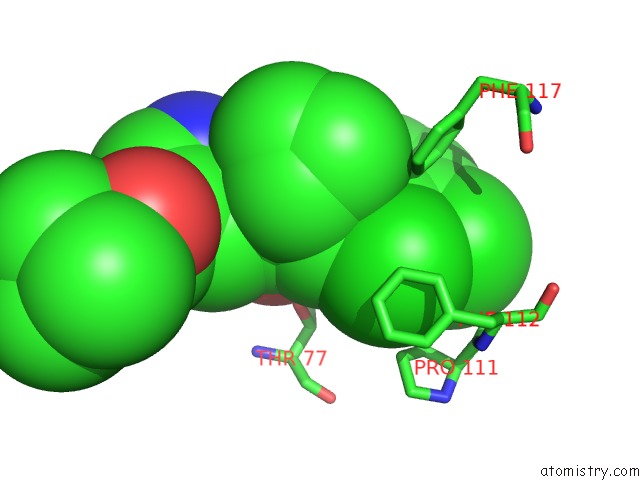
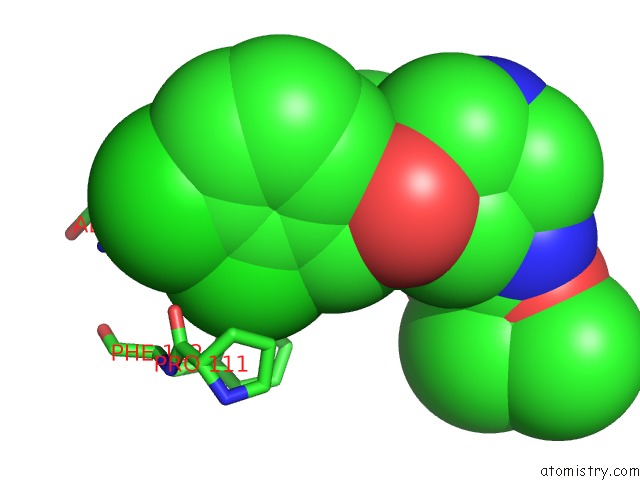
Chlorine binding site 1 out of 4 in 4pyv
Go back to
Chlorine binding site 1 out
of 4 in the Crystal Structure of Renin in Complex with COMPOUND4
Mono view
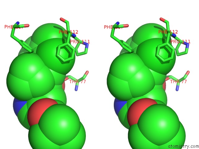
Stereo pair view
Mono view
Stereo pair view
A full contact list of Chlorine with other atoms in the Cl binding
site number 1 of Crystal Structure of Renin in Complex with COMPOUND4 within 5.0Å range:
|
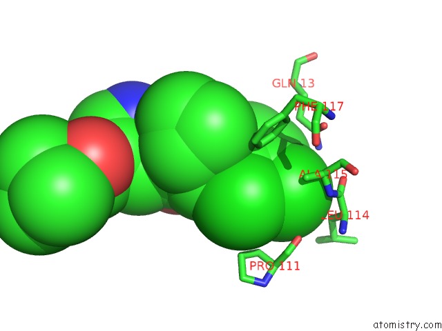
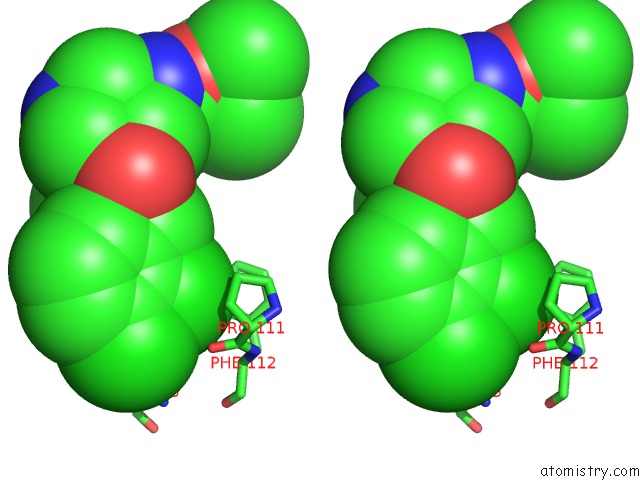
Chlorine binding site 2 out of 4 in 4pyv
Go back to
Chlorine binding site 2 out
of 4 in the Crystal Structure of Renin in Complex with COMPOUND4
Mono view
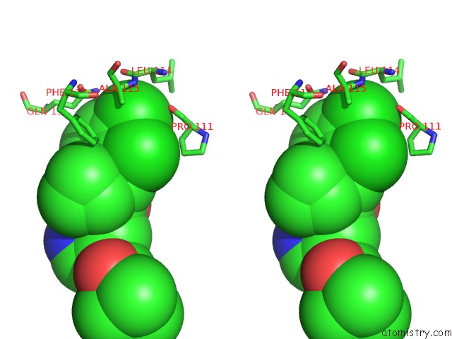
Stereo pair view
Mono view
Stereo pair view
A full contact list of Chlorine with other atoms in the Cl binding
site number 2 of Crystal Structure of Renin in Complex with COMPOUND4 within 5.0Å range:
|
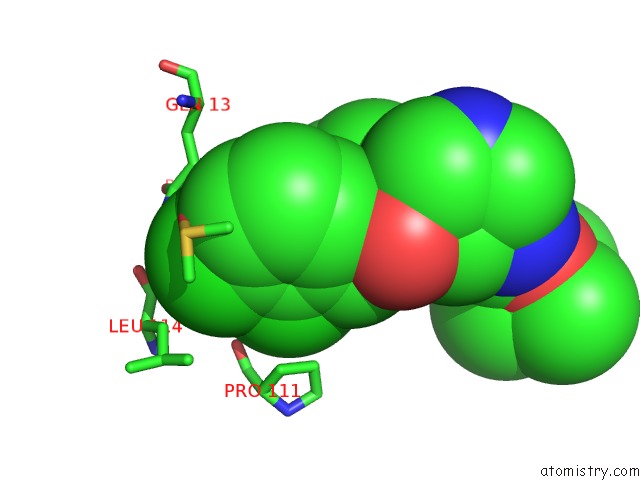
Chlorine binding site 3 out of 4 in 4pyv
Go back to
Chlorine binding site 3 out
of 4 in the Crystal Structure of Renin in Complex with COMPOUND4
Mono view
Stereo pair view
Mono view
Stereo pair view
A full contact list of Chlorine with other atoms in the Cl binding
site number 3 of Crystal Structure of Renin in Complex with COMPOUND4 within 5.0Å range:
|
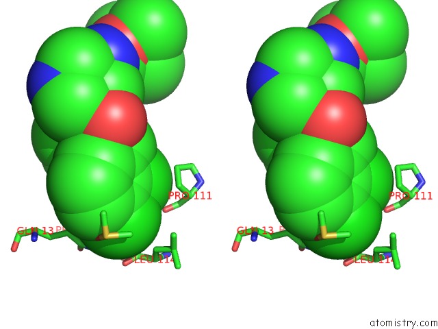
Chlorine binding site 4 out of 4 in 4pyv
Go back to
Chlorine binding site 4 out
of 4 in the Crystal Structure of Renin in Complex with COMPOUND4
Mono view
Stereo pair view
Mono view
Stereo pair view
A full contact list of Chlorine with other atoms in the Cl binding
site number 4 of Crystal Structure of Renin in Complex with COMPOUND4 within 5.0Å range:
|
Reference:
T.Ehara,
O.Irie,
T.Kosaka,
T.Kanazawa,
W.Breitenstein,
P.Grosche,
N.Ostermann,
M.Suzuki,
S.Kawakami,
K.Konishi,
Y.Hitomi,
A.Toyao,
H.Gunji,
F.Cumin,
N.Schiering,
T.Wagner,
D.F.Rigel,
R.L.Webb,
J.Maibaum,
F.Yokokawa.
Structure-Based Design of Substituted Piperidines As A New Class of Highly Efficacious Oral Direct Renin Inhibitors. Acs Med Chem Lett V. 5 787 2014.
PubMed: 25050166
DOI: 10.1021/ML500137B
Page generated: Fri Jul 26 00:13:53 2024
PubMed: 25050166
DOI: 10.1021/ML500137B
Last articles
Zn in 9J0NZn in 9J0O
Zn in 9J0P
Zn in 9FJX
Zn in 9EKB
Zn in 9C0F
Zn in 9CAH
Zn in 9CH0
Zn in 9CH3
Zn in 9CH1