Chlorine »
PDB 4px1-4q63 »
4q4x »
Chlorine in PDB 4q4x: Crystal Structure of Coxsackievirus A24V Soaked with 6'-Sialyllactose (6SL)
Protein crystallography data
The structure of Crystal Structure of Coxsackievirus A24V Soaked with 6'-Sialyllactose (6SL), PDB code: 4q4x
was solved by
G.Zocher,
T.Stehle,
with X-Ray Crystallography technique. A brief refinement statistics is given in the table below:
| Resolution Low / High (Å) | 49.95 / 1.65 |
| Space group | I 2 2 2 |
| Cell size a, b, c (Å), α, β, γ (°) | 306.398, 366.096, 367.700, 90.00, 90.00, 90.00 |
| R / Rfree (%) | 14.9 / n/a |
Other elements in 4q4x:
The structure of Crystal Structure of Coxsackievirus A24V Soaked with 6'-Sialyllactose (6SL) also contains other interesting chemical elements:
| Calcium | (Ca) | 5 atoms |
| Sodium | (Na) | 1 atom |
Chlorine Binding Sites:
The binding sites of Chlorine atom in the Crystal Structure of Coxsackievirus A24V Soaked with 6'-Sialyllactose (6SL)
(pdb code 4q4x). This binding sites where shown within
5.0 Angstroms radius around Chlorine atom.
In total 5 binding sites of Chlorine where determined in the Crystal Structure of Coxsackievirus A24V Soaked with 6'-Sialyllactose (6SL), PDB code: 4q4x:
Jump to Chlorine binding site number: 1; 2; 3; 4; 5;
In total 5 binding sites of Chlorine where determined in the Crystal Structure of Coxsackievirus A24V Soaked with 6'-Sialyllactose (6SL), PDB code: 4q4x:
Jump to Chlorine binding site number: 1; 2; 3; 4; 5;
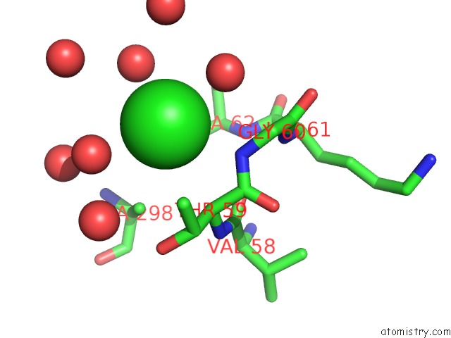
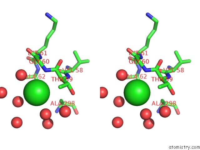
Chlorine binding site 1 out of 5 in 4q4x
Go back to
Chlorine binding site 1 out
of 5 in the Crystal Structure of Coxsackievirus A24V Soaked with 6'-Sialyllactose (6SL)

Mono view
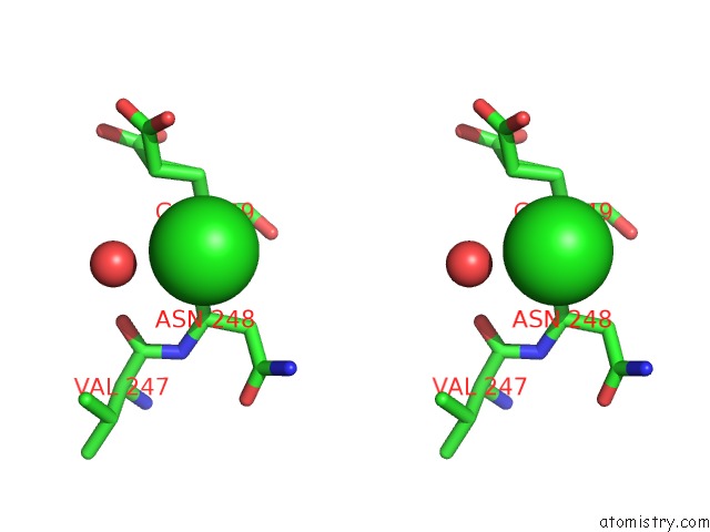
Stereo pair view
Mono view
Stereo pair view
A full contact list of Chlorine with other atoms in the Cl binding
site number 1 of Crystal Structure of Coxsackievirus A24V Soaked with 6'-Sialyllactose (6SL) within 5.0Å range:
|
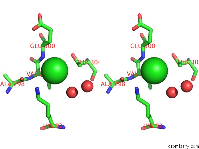
Chlorine binding site 2 out of 5 in 4q4x
Go back to
Chlorine binding site 2 out
of 5 in the Crystal Structure of Coxsackievirus A24V Soaked with 6'-Sialyllactose (6SL)

Mono view

Stereo pair view
Mono view
Stereo pair view
A full contact list of Chlorine with other atoms in the Cl binding
site number 2 of Crystal Structure of Coxsackievirus A24V Soaked with 6'-Sialyllactose (6SL) within 5.0Å range:
|
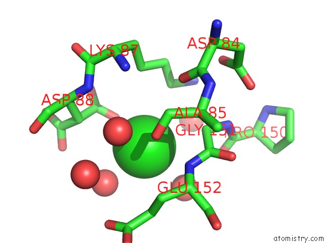
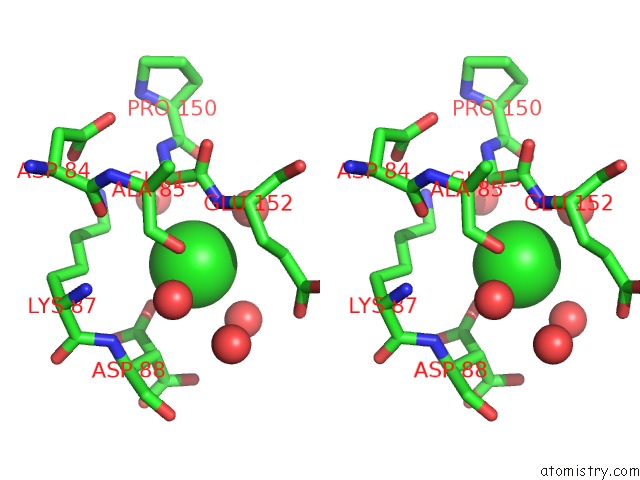
Chlorine binding site 3 out of 5 in 4q4x
Go back to
Chlorine binding site 3 out
of 5 in the Crystal Structure of Coxsackievirus A24V Soaked with 6'-Sialyllactose (6SL)

Mono view
Stereo pair view
Mono view
Stereo pair view
A full contact list of Chlorine with other atoms in the Cl binding
site number 3 of Crystal Structure of Coxsackievirus A24V Soaked with 6'-Sialyllactose (6SL) within 5.0Å range:
|
Chlorine binding site 4 out of 5 in 4q4x
Go back to
Chlorine binding site 4 out
of 5 in the Crystal Structure of Coxsackievirus A24V Soaked with 6'-Sialyllactose (6SL)
Mono view
Stereo pair view
Mono view
Stereo pair view
A full contact list of Chlorine with other atoms in the Cl binding
site number 4 of Crystal Structure of Coxsackievirus A24V Soaked with 6'-Sialyllactose (6SL) within 5.0Å range:
|
Chlorine binding site 5 out of 5 in 4q4x
Go back to
Chlorine binding site 5 out
of 5 in the Crystal Structure of Coxsackievirus A24V Soaked with 6'-Sialyllactose (6SL)
Mono view
Stereo pair view
Mono view
Stereo pair view
A full contact list of Chlorine with other atoms in the Cl binding
site number 5 of Crystal Structure of Coxsackievirus A24V Soaked with 6'-Sialyllactose (6SL) within 5.0Å range:
|
Reference:
G.Zocher,
N.Mistry,
M.Frank,
I.Hahnlein-Schick,
J.O.Ekstrom,
N.Arnberg,
T.Stehle.
A Sialic Acid Binding Site in A Human Picornavirus. Plos Pathog. V. 10 04401 2014.
ISSN: ISSN 1553-7366
PubMed: 25329320
DOI: 10.1371/JOURNAL.PPAT.1004401
Page generated: Fri Jul 26 00:18:28 2024
ISSN: ISSN 1553-7366
PubMed: 25329320
DOI: 10.1371/JOURNAL.PPAT.1004401
Last articles
Zn in 9MJ5Zn in 9HNW
Zn in 9G0L
Zn in 9FNE
Zn in 9DZN
Zn in 9E0I
Zn in 9D32
Zn in 9DAK
Zn in 8ZXC
Zn in 8ZUF