Chlorine »
PDB 4rjt-4rsd »
4rqk »
Chlorine in PDB 4rqk: Crystal Structure of PDK1 in Complex with Atp and the Pif-Pocket Ligand RS1
Enzymatic activity of Crystal Structure of PDK1 in Complex with Atp and the Pif-Pocket Ligand RS1
All present enzymatic activity of Crystal Structure of PDK1 in Complex with Atp and the Pif-Pocket Ligand RS1:
2.7.11.1;
2.7.11.1;
Protein crystallography data
The structure of Crystal Structure of PDK1 in Complex with Atp and the Pif-Pocket Ligand RS1, PDB code: 4rqk
was solved by
T.J.Rettenmaier,
J.A.Wells,
with X-Ray Crystallography technique. A brief refinement statistics is given in the table below:
| Resolution Low / High (Å) | 46.69 / 1.55 |
| Space group | C 1 2 1 |
| Cell size a, b, c (Å), α, β, γ (°) | 148.440, 44.239, 47.496, 90.00, 100.58, 90.00 |
| R / Rfree (%) | 13.1 / 16.7 |
Chlorine Binding Sites:
The binding sites of Chlorine atom in the Crystal Structure of PDK1 in Complex with Atp and the Pif-Pocket Ligand RS1
(pdb code 4rqk). This binding sites where shown within
5.0 Angstroms radius around Chlorine atom.
In total only one binding site of Chlorine was determined in the Crystal Structure of PDK1 in Complex with Atp and the Pif-Pocket Ligand RS1, PDB code: 4rqk:
In total only one binding site of Chlorine was determined in the Crystal Structure of PDK1 in Complex with Atp and the Pif-Pocket Ligand RS1, PDB code: 4rqk:
Chlorine binding site 1 out of 1 in 4rqk
Go back to
Chlorine binding site 1 out
of 1 in the Crystal Structure of PDK1 in Complex with Atp and the Pif-Pocket Ligand RS1
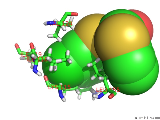
Mono view
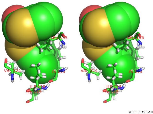
Stereo pair view
Mono view
Stereo pair view
A full contact list of Chlorine with other atoms in the Cl binding
site number 1 of Crystal Structure of PDK1 in Complex with Atp and the Pif-Pocket Ligand RS1 within 5.0Å range:
|
Reference:
T.J.Rettenmaier,
J.D.Sadowsky,
N.D.Thomsen,
S.C.Chen,
A.K.Doak,
M.R.Arkin,
J.A.Wells.
A Small-Molecule Mimic of A Peptide Docking Motif Inhibits the Protein Kinase PDK1 Proc.Natl.Acad.Sci.Usa 2014.
ISSN: ESSN 1091-6490
Page generated: Fri Jul 26 01:26:23 2024
ISSN: ESSN 1091-6490
Last articles
Zn in 9J0NZn in 9J0O
Zn in 9J0P
Zn in 9FJX
Zn in 9EKB
Zn in 9C0F
Zn in 9CAH
Zn in 9CH0
Zn in 9CH3
Zn in 9CH1