Chlorine »
PDB 4rjt-4rsd »
4rqo »
Chlorine in PDB 4rqo: Crystal Structure of L-Serine Dehydratase From Legionella Pneumophila
Enzymatic activity of Crystal Structure of L-Serine Dehydratase From Legionella Pneumophila
All present enzymatic activity of Crystal Structure of L-Serine Dehydratase From Legionella Pneumophila:
4.3.1.17;
4.3.1.17;
Protein crystallography data
The structure of Crystal Structure of L-Serine Dehydratase From Legionella Pneumophila, PDB code: 4rqo
was solved by
J.B.Thoden,
H.M.Holden,
G.A.Grant,
with X-Ray Crystallography technique. A brief refinement statistics is given in the table below:
| Resolution Low / High (Å) | 29.45 / 2.25 |
| Space group | P 31 2 1 |
| Cell size a, b, c (Å), α, β, γ (°) | 81.390, 81.390, 267.521, 90.00, 90.00, 120.00 |
| R / Rfree (%) | 19.8 / 25.8 |
Other elements in 4rqo:
The structure of Crystal Structure of L-Serine Dehydratase From Legionella Pneumophila also contains other interesting chemical elements:
| Iron | (Fe) | 8 atoms |
Chlorine Binding Sites:
The binding sites of Chlorine atom in the Crystal Structure of L-Serine Dehydratase From Legionella Pneumophila
(pdb code 4rqo). This binding sites where shown within
5.0 Angstroms radius around Chlorine atom.
In total 2 binding sites of Chlorine where determined in the Crystal Structure of L-Serine Dehydratase From Legionella Pneumophila, PDB code: 4rqo:
Jump to Chlorine binding site number: 1; 2;
In total 2 binding sites of Chlorine where determined in the Crystal Structure of L-Serine Dehydratase From Legionella Pneumophila, PDB code: 4rqo:
Jump to Chlorine binding site number: 1; 2;
Chlorine binding site 1 out of 2 in 4rqo
Go back to
Chlorine binding site 1 out
of 2 in the Crystal Structure of L-Serine Dehydratase From Legionella Pneumophila
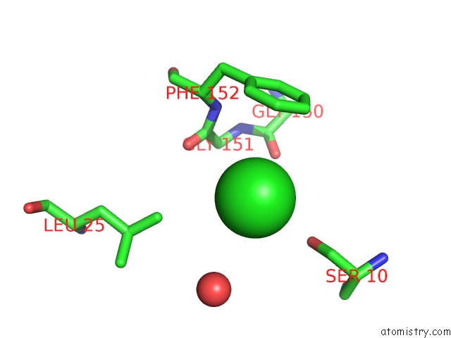
Mono view
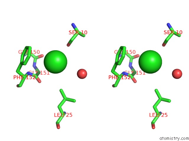
Stereo pair view
Mono view
Stereo pair view
A full contact list of Chlorine with other atoms in the Cl binding
site number 1 of Crystal Structure of L-Serine Dehydratase From Legionella Pneumophila within 5.0Å range:
|
Chlorine binding site 2 out of 2 in 4rqo
Go back to
Chlorine binding site 2 out
of 2 in the Crystal Structure of L-Serine Dehydratase From Legionella Pneumophila
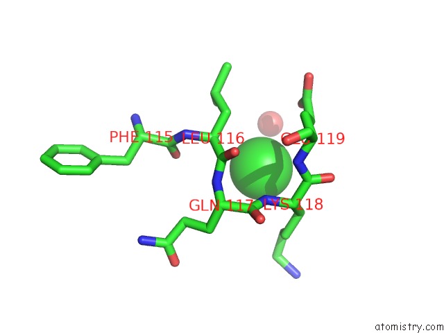
Mono view
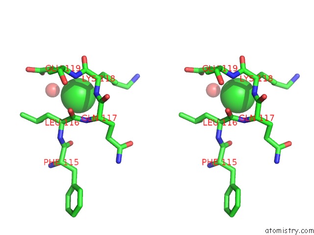
Stereo pair view
Mono view
Stereo pair view
A full contact list of Chlorine with other atoms in the Cl binding
site number 2 of Crystal Structure of L-Serine Dehydratase From Legionella Pneumophila within 5.0Å range:
|
Reference:
J.B.Thoden,
H.M.Holden,
G.A.Grant.
Structure of L-Serine Dehydratase From Legionella Pneumophila: Novel Use of the C-Terminal Cysteine As An Intrinsic Competitive Inhibitor. Biochemistry 2014.
ISSN: ISSN 0006-2960
PubMed: 25380533
DOI: 10.1021/BI501253W
Page generated: Fri Jul 26 01:26:25 2024
ISSN: ISSN 0006-2960
PubMed: 25380533
DOI: 10.1021/BI501253W
Last articles
Zn in 9J0NZn in 9J0O
Zn in 9J0P
Zn in 9FJX
Zn in 9EKB
Zn in 9C0F
Zn in 9CAH
Zn in 9CH0
Zn in 9CH3
Zn in 9CH1