Chlorine »
PDB 4s28-4tsq »
4tlh »
Chlorine in PDB 4tlh: Monoclinic Crystal Structure of Eutl From Clostridium Perfringens
Protein crystallography data
The structure of Monoclinic Crystal Structure of Eutl From Clostridium Perfringens, PDB code: 4tlh
was solved by
M.C.Thompson,
T.O.Yeates,
with X-Ray Crystallography technique. A brief refinement statistics is given in the table below:
| Resolution Low / High (Å) | 33.43 / 1.70 |
| Space group | C 1 2 1 |
| Cell size a, b, c (Å), α, β, γ (°) | 118.440, 66.030, 79.380, 90.00, 108.27, 90.00 |
| R / Rfree (%) | 16.3 / 19.5 |
Other elements in 4tlh:
The structure of Monoclinic Crystal Structure of Eutl From Clostridium Perfringens also contains other interesting chemical elements:
| Sodium | (Na) | 2 atoms |
Chlorine Binding Sites:
The binding sites of Chlorine atom in the Monoclinic Crystal Structure of Eutl From Clostridium Perfringens
(pdb code 4tlh). This binding sites where shown within
5.0 Angstroms radius around Chlorine atom.
In total only one binding site of Chlorine was determined in the Monoclinic Crystal Structure of Eutl From Clostridium Perfringens, PDB code: 4tlh:
In total only one binding site of Chlorine was determined in the Monoclinic Crystal Structure of Eutl From Clostridium Perfringens, PDB code: 4tlh:
Chlorine binding site 1 out of 1 in 4tlh
Go back to
Chlorine binding site 1 out
of 1 in the Monoclinic Crystal Structure of Eutl From Clostridium Perfringens
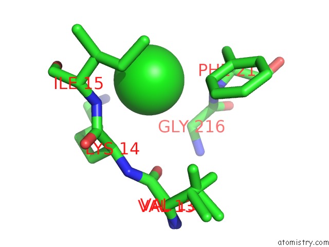
Mono view
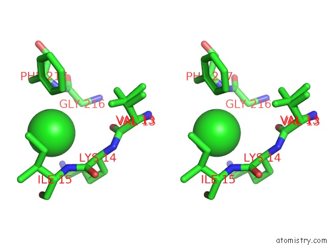
Stereo pair view
Mono view
Stereo pair view
A full contact list of Chlorine with other atoms in the Cl binding
site number 1 of Monoclinic Crystal Structure of Eutl From Clostridium Perfringens within 5.0Å range:
|
Reference:
M.C.Thompson,
D.Cascio,
T.O.Yeates.
Microfocus Diffraction From Different Regions of A Protein Crystal: Structural Variations and Unit-Cell Polymorphism Acta Crystallogr.,Sect.D 2018.
ISSN: ESSN 1399-0047
DOI: 10.1107/S2059798318003479
Page generated: Fri Jul 26 01:46:22 2024
ISSN: ESSN 1399-0047
DOI: 10.1107/S2059798318003479
Last articles
Zn in 9MJ5Zn in 9HNW
Zn in 9G0L
Zn in 9FNE
Zn in 9DZN
Zn in 9E0I
Zn in 9D32
Zn in 9DAK
Zn in 8ZXC
Zn in 8ZUF