Chlorine »
PDB 4s28-4tsq »
4tm7 »
Chlorine in PDB 4tm7: Crystal Structure of 6-Phosphogluconolactonase From Mycobacterium Smegmatis N131D Mutant Soaked with CUSO4
Enzymatic activity of Crystal Structure of 6-Phosphogluconolactonase From Mycobacterium Smegmatis N131D Mutant Soaked with CUSO4
All present enzymatic activity of Crystal Structure of 6-Phosphogluconolactonase From Mycobacterium Smegmatis N131D Mutant Soaked with CUSO4:
3.1.1.31;
3.1.1.31;
Protein crystallography data
The structure of Crystal Structure of 6-Phosphogluconolactonase From Mycobacterium Smegmatis N131D Mutant Soaked with CUSO4, PDB code: 4tm7
was solved by
N.Fujieda,
E.Stuttfeld,
T.Maier,
with X-Ray Crystallography technique. A brief refinement statistics is given in the table below:
| Resolution Low / High (Å) | 74.23 / 1.39 |
| Space group | P 32 1 2 |
| Cell size a, b, c (Å), α, β, γ (°) | 42.520, 42.520, 222.700, 90.00, 90.00, 120.00 |
| R / Rfree (%) | 14.9 / 17.8 |
Other elements in 4tm7:
The structure of Crystal Structure of 6-Phosphogluconolactonase From Mycobacterium Smegmatis N131D Mutant Soaked with CUSO4 also contains other interesting chemical elements:
| Copper | (Cu) | 16 atoms |
Chlorine Binding Sites:
The binding sites of Chlorine atom in the Crystal Structure of 6-Phosphogluconolactonase From Mycobacterium Smegmatis N131D Mutant Soaked with CUSO4
(pdb code 4tm7). This binding sites where shown within
5.0 Angstroms radius around Chlorine atom.
In total 2 binding sites of Chlorine where determined in the Crystal Structure of 6-Phosphogluconolactonase From Mycobacterium Smegmatis N131D Mutant Soaked with CUSO4, PDB code: 4tm7:
Jump to Chlorine binding site number: 1; 2;
In total 2 binding sites of Chlorine where determined in the Crystal Structure of 6-Phosphogluconolactonase From Mycobacterium Smegmatis N131D Mutant Soaked with CUSO4, PDB code: 4tm7:
Jump to Chlorine binding site number: 1; 2;
Chlorine binding site 1 out of 2 in 4tm7
Go back to
Chlorine binding site 1 out
of 2 in the Crystal Structure of 6-Phosphogluconolactonase From Mycobacterium Smegmatis N131D Mutant Soaked with CUSO4
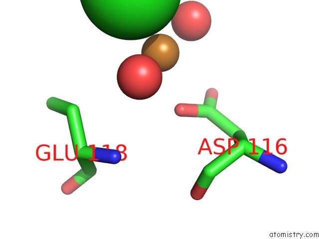
Mono view
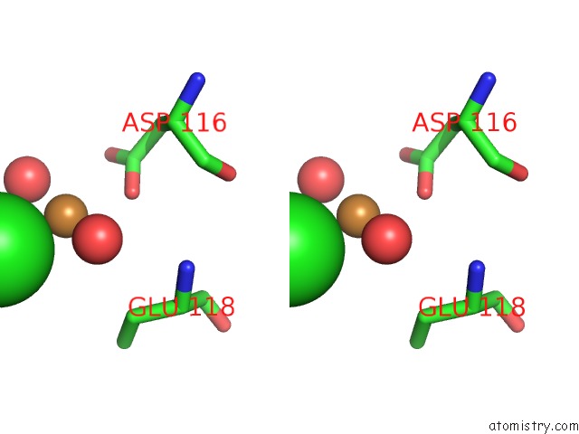
Stereo pair view
Mono view
Stereo pair view
A full contact list of Chlorine with other atoms in the Cl binding
site number 1 of Crystal Structure of 6-Phosphogluconolactonase From Mycobacterium Smegmatis N131D Mutant Soaked with CUSO4 within 5.0Å range:
|
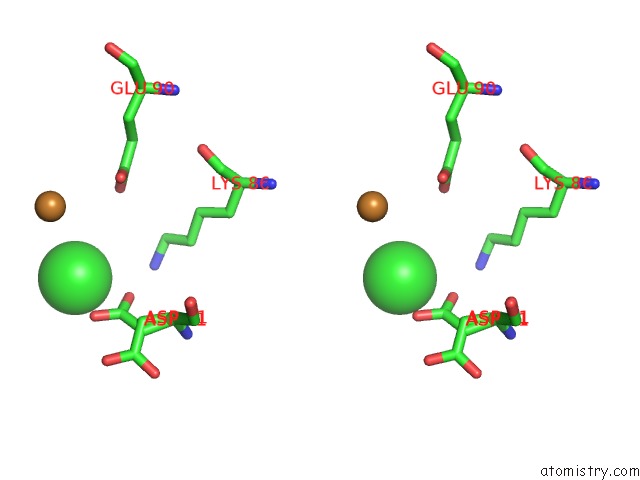
Chlorine binding site 2 out of 2 in 4tm7
Go back to
Chlorine binding site 2 out
of 2 in the Crystal Structure of 6-Phosphogluconolactonase From Mycobacterium Smegmatis N131D Mutant Soaked with CUSO4
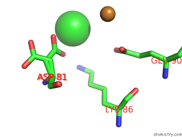
Mono view
Stereo pair view
Mono view
Stereo pair view
A full contact list of Chlorine with other atoms in the Cl binding
site number 2 of Crystal Structure of 6-Phosphogluconolactonase From Mycobacterium Smegmatis N131D Mutant Soaked with CUSO4 within 5.0Å range:
|
Reference:
N.Fujieda,
J.Schatti,
E.Stuttfeld,
K.Ohkubo,
T.Maier,
S.Fukuzumi,
T.R.Ward.
Enzyme Repurposing of A Hydrolase As An Emergent Peroxidase Upon Metal Binding. Chem Sci V. 6 4060 2015.
ISSN: ISSN 2041-6520
PubMed: 29218172
DOI: 10.1039/C5SC01065A
Page generated: Fri Jul 11 21:44:28 2025
ISSN: ISSN 2041-6520
PubMed: 29218172
DOI: 10.1039/C5SC01065A
Last articles
F in 4ITJF in 4ISF
F in 4IKS
F in 4ISE
F in 4IQV
F in 4IQW
F in 4IQT
F in 4IQU
F in 4INB
F in 4IKT