Chlorine »
PDB 4uij-4uvn »
4uut »
Chlorine in PDB 4uut: Crystal Structure of the Ultrabithorax Protein
Protein crystallography data
The structure of Crystal Structure of the Ultrabithorax Protein, PDB code: 4uut
was solved by
N.Foos,
M.J.Mate,
M.Ortiz-Lombardia,
with X-Ray Crystallography technique. A brief refinement statistics is given in the table below:
| Resolution Low / High (Å) | 69.75 / 2.80 |
| Space group | P 41 3 2 |
| Cell size a, b, c (Å), α, β, γ (°) | 98.648, 98.648, 98.648, 90.00, 90.00, 90.00 |
| R / Rfree (%) | 21.75 / 23.66 |
Chlorine Binding Sites:
The binding sites of Chlorine atom in the Crystal Structure of the Ultrabithorax Protein
(pdb code 4uut). This binding sites where shown within
5.0 Angstroms radius around Chlorine atom.
In total 2 binding sites of Chlorine where determined in the Crystal Structure of the Ultrabithorax Protein, PDB code: 4uut:
Jump to Chlorine binding site number: 1; 2;
In total 2 binding sites of Chlorine where determined in the Crystal Structure of the Ultrabithorax Protein, PDB code: 4uut:
Jump to Chlorine binding site number: 1; 2;
Chlorine binding site 1 out of 2 in 4uut
Go back to
Chlorine binding site 1 out
of 2 in the Crystal Structure of the Ultrabithorax Protein
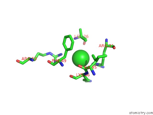
Mono view
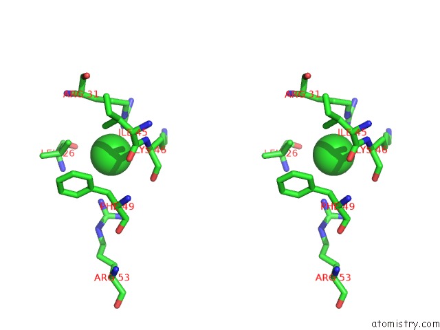
Stereo pair view
Mono view
Stereo pair view
A full contact list of Chlorine with other atoms in the Cl binding
site number 1 of Crystal Structure of the Ultrabithorax Protein within 5.0Å range:
|
Chlorine binding site 2 out of 2 in 4uut
Go back to
Chlorine binding site 2 out
of 2 in the Crystal Structure of the Ultrabithorax Protein
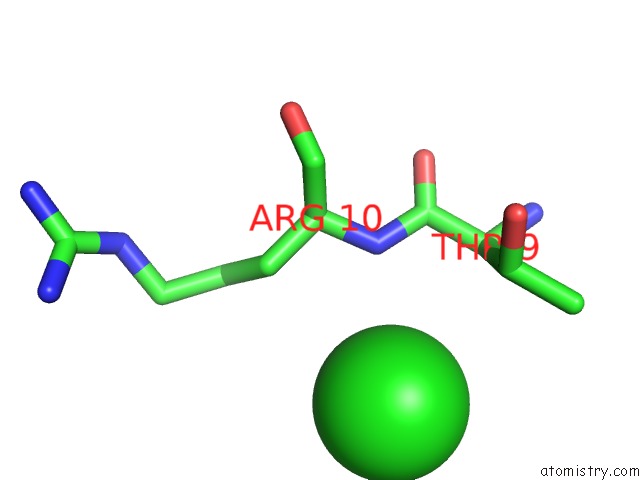
Mono view
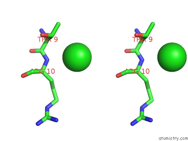
Stereo pair view
Mono view
Stereo pair view
A full contact list of Chlorine with other atoms in the Cl binding
site number 2 of Crystal Structure of the Ultrabithorax Protein within 5.0Å range:
|
Reference:
N.Foos,
C.Maurel-Zaffran,
M.J.Mate,
R.Vincentelli,
M.Hainaut,
H.Berenger,
J.Pradel,
A.J.Saurin,
M.Ortiz-Lombardia,
Y.Graba.
A Flexible Extension of the Drosophila Ultrabithorax Homeodomain Defines A Novel Hox/Pbc Interaction Mode. Structure V. 23 270 2015.
ISSN: ISSN 0969-2126
PubMed: 25651060
DOI: 10.1016/J.STR.2014.12.011
Page generated: Fri Jul 26 02:32:19 2024
ISSN: ISSN 0969-2126
PubMed: 25651060
DOI: 10.1016/J.STR.2014.12.011
Last articles
Zn in 9J0NZn in 9J0O
Zn in 9J0P
Zn in 9FJX
Zn in 9EKB
Zn in 9C0F
Zn in 9CAH
Zn in 9CH0
Zn in 9CH3
Zn in 9CH1