Chlorine »
PDB 4wl6-4ws6 »
4wl6 »
Chlorine in PDB 4wl6: Raster-Scanning Protein Crystallography Using Micro and Nano-Focused Synchrotron Beams
Enzymatic activity of Raster-Scanning Protein Crystallography Using Micro and Nano-Focused Synchrotron Beams
All present enzymatic activity of Raster-Scanning Protein Crystallography Using Micro and Nano-Focused Synchrotron Beams:
3.2.1.17;
3.2.1.17;
Protein crystallography data
The structure of Raster-Scanning Protein Crystallography Using Micro and Nano-Focused Synchrotron Beams, PDB code: 4wl6
was solved by
N.Coquelle,
U.Kapp,
A.Shilova,
B.Weinhausen,
M.Burghammer,
J.P.Colletier,
with X-Ray Crystallography technique. A brief refinement statistics is given in the table below:
| Resolution Low / High (Å) | 39.00 / 1.85 |
| Space group | P 43 21 2 |
| Cell size a, b, c (Å), α, β, γ (°) | 77.990, 77.990, 38.510, 90.00, 90.00, 90.00 |
| R / Rfree (%) | 22.8 / 27.1 |
Chlorine Binding Sites:
The binding sites of Chlorine atom in the Raster-Scanning Protein Crystallography Using Micro and Nano-Focused Synchrotron Beams
(pdb code 4wl6). This binding sites where shown within
5.0 Angstroms radius around Chlorine atom.
In total only one binding site of Chlorine was determined in the Raster-Scanning Protein Crystallography Using Micro and Nano-Focused Synchrotron Beams, PDB code: 4wl6:
In total only one binding site of Chlorine was determined in the Raster-Scanning Protein Crystallography Using Micro and Nano-Focused Synchrotron Beams, PDB code: 4wl6:
Chlorine binding site 1 out of 1 in 4wl6
Go back to
Chlorine binding site 1 out
of 1 in the Raster-Scanning Protein Crystallography Using Micro and Nano-Focused Synchrotron Beams
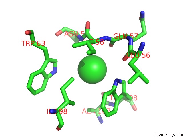
Mono view
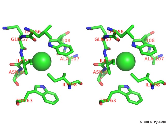
Stereo pair view
Mono view
Stereo pair view
A full contact list of Chlorine with other atoms in the Cl binding
site number 1 of Raster-Scanning Protein Crystallography Using Micro and Nano-Focused Synchrotron Beams within 5.0Å range:
|
Reference:
N.Coquelle,
A.S.Brewster,
U.Kapp,
A.Shilova,
B.Weimhausen,
M.Burghammer,
J.-P.Colletier.
Raster-Scanning Protein Crystallography Using Micro and Nano-Focused Synchrotron Beams. Acta Crystallogr.,Sect.D V. 71 1184 2015.
ISSN: ESSN 1399-0047
DOI: 10.1107/S1399004715004514
Page generated: Fri Jul 11 22:33:09 2025
ISSN: ESSN 1399-0047
DOI: 10.1107/S1399004715004514
Last articles
Fe in 2YXOFe in 2YRS
Fe in 2YXC
Fe in 2YNM
Fe in 2YVJ
Fe in 2YP1
Fe in 2YU2
Fe in 2YU1
Fe in 2YQB
Fe in 2YOO