Chlorine »
PDB 4ws7-4wxe »
4wt9 »
Chlorine in PDB 4wt9: Apo Crystal Structure of Hcv NS5B Genotype 2A Jfh-1 Isolate with E86Q E87Q S15G C223H V321I and DELTA8 Mutations
Enzymatic activity of Apo Crystal Structure of Hcv NS5B Genotype 2A Jfh-1 Isolate with E86Q E87Q S15G C223H V321I and DELTA8 Mutations
All present enzymatic activity of Apo Crystal Structure of Hcv NS5B Genotype 2A Jfh-1 Isolate with E86Q E87Q S15G C223H V321I and DELTA8 Mutations:
2.7.7.48;
2.7.7.48;
Protein crystallography data
The structure of Apo Crystal Structure of Hcv NS5B Genotype 2A Jfh-1 Isolate with E86Q E87Q S15G C223H V321I and DELTA8 Mutations, PDB code: 4wt9
was solved by
T.E.Edwards,
T.C.Appleby,
with X-Ray Crystallography technique. A brief refinement statistics is given in the table below:
| Resolution Low / High (Å) | 50.00 / 2.50 |
| Space group | P 65 |
| Cell size a, b, c (Å), α, β, γ (°) | 140.370, 140.370, 92.230, 90.00, 90.00, 120.00 |
| R / Rfree (%) | 19.3 / 24.5 |
Chlorine Binding Sites:
The binding sites of Chlorine atom in the Apo Crystal Structure of Hcv NS5B Genotype 2A Jfh-1 Isolate with E86Q E87Q S15G C223H V321I and DELTA8 Mutations
(pdb code 4wt9). This binding sites where shown within
5.0 Angstroms radius around Chlorine atom.
In total only one binding site of Chlorine was determined in the Apo Crystal Structure of Hcv NS5B Genotype 2A Jfh-1 Isolate with E86Q E87Q S15G C223H V321I and DELTA8 Mutations, PDB code: 4wt9:
In total only one binding site of Chlorine was determined in the Apo Crystal Structure of Hcv NS5B Genotype 2A Jfh-1 Isolate with E86Q E87Q S15G C223H V321I and DELTA8 Mutations, PDB code: 4wt9:
Chlorine binding site 1 out of 1 in 4wt9
Go back to
Chlorine binding site 1 out
of 1 in the Apo Crystal Structure of Hcv NS5B Genotype 2A Jfh-1 Isolate with E86Q E87Q S15G C223H V321I and DELTA8 Mutations
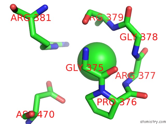
Mono view
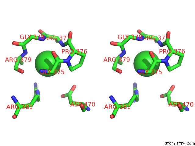
Stereo pair view
Mono view
Stereo pair view
A full contact list of Chlorine with other atoms in the Cl binding
site number 1 of Apo Crystal Structure of Hcv NS5B Genotype 2A Jfh-1 Isolate with E86Q E87Q S15G C223H V321I and DELTA8 Mutations within 5.0Å range:
|
Reference:
T.C.Appleby,
J.Perry,
E.Murakami,
O.Barauskas,
J.Feng,
A.Cho,
D.Fox Iii,
D.R.Wetmore,
M.E.Mcgrath,
A.S.Ray,
M.J.Sofia,
S.Swaminathan,
T.E.Edwards.
Structural Basis For Rna Replication By the Hepatitis C Virus Polymerase To Be Published.
Page generated: Fri Jul 11 22:38:45 2025
Last articles
Fe in 2YXOFe in 2YRS
Fe in 2YXC
Fe in 2YNM
Fe in 2YVJ
Fe in 2YP1
Fe in 2YU2
Fe in 2YU1
Fe in 2YQB
Fe in 2YOO