Chlorine »
PDB 4wxp-4x97 »
4x1x »
Chlorine in PDB 4x1x: Crystal Structure of Rhdvb P Domain in Complex with Lewis Y
Protein crystallography data
The structure of Crystal Structure of Rhdvb P Domain in Complex with Lewis Y, PDB code: 4x1x
was solved by
M.M.Leuthold,
G.S.Hansman,
with X-Ray Crystallography technique. A brief refinement statistics is given in the table below:
| Resolution Low / High (Å) | 48.22 / 1.60 |
| Space group | P 1 21 1 |
| Cell size a, b, c (Å), α, β, γ (°) | 59.440, 84.150, 62.660, 90.00, 110.11, 90.00 |
| R / Rfree (%) | 15.5 / 18.5 |
Chlorine Binding Sites:
The binding sites of Chlorine atom in the Crystal Structure of Rhdvb P Domain in Complex with Lewis Y
(pdb code 4x1x). This binding sites where shown within
5.0 Angstroms radius around Chlorine atom.
In total 3 binding sites of Chlorine where determined in the Crystal Structure of Rhdvb P Domain in Complex with Lewis Y, PDB code: 4x1x:
Jump to Chlorine binding site number: 1; 2; 3;
In total 3 binding sites of Chlorine where determined in the Crystal Structure of Rhdvb P Domain in Complex with Lewis Y, PDB code: 4x1x:
Jump to Chlorine binding site number: 1; 2; 3;
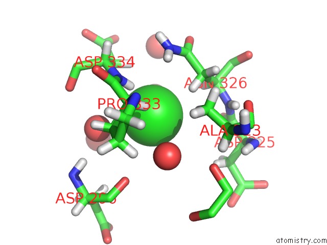
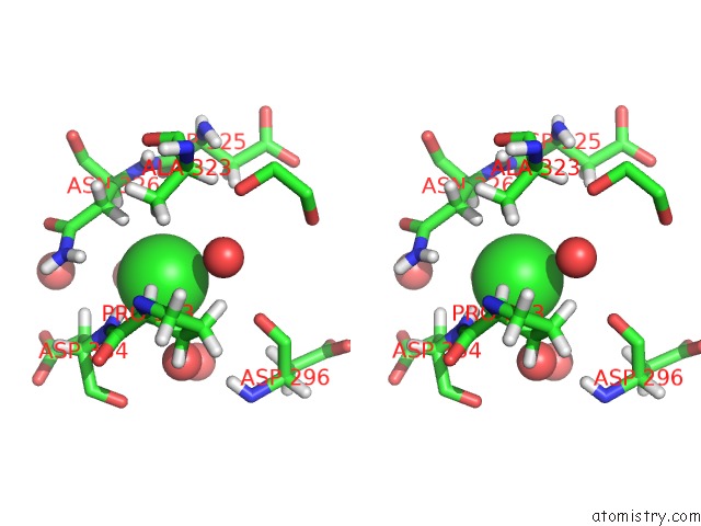
Chlorine binding site 1 out of 3 in 4x1x
Go back to
Chlorine binding site 1 out
of 3 in the Crystal Structure of Rhdvb P Domain in Complex with Lewis Y
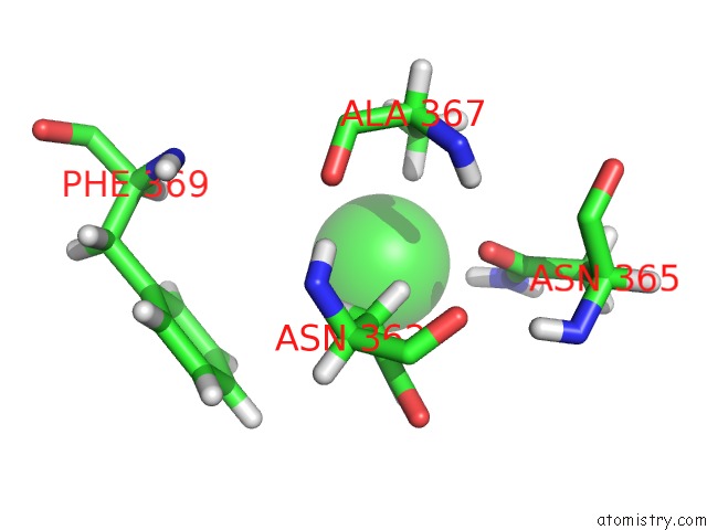
Mono view
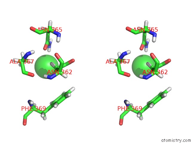
Stereo pair view
Mono view
Stereo pair view
A full contact list of Chlorine with other atoms in the Cl binding
site number 1 of Crystal Structure of Rhdvb P Domain in Complex with Lewis Y within 5.0Å range:
|
Chlorine binding site 2 out of 3 in 4x1x
Go back to
Chlorine binding site 2 out
of 3 in the Crystal Structure of Rhdvb P Domain in Complex with Lewis Y
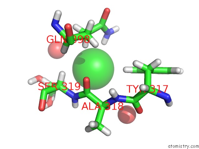
Mono view
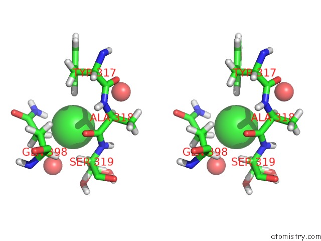
Stereo pair view
Mono view
Stereo pair view
A full contact list of Chlorine with other atoms in the Cl binding
site number 2 of Crystal Structure of Rhdvb P Domain in Complex with Lewis Y within 5.0Å range:
|
Chlorine binding site 3 out of 3 in 4x1x
Go back to
Chlorine binding site 3 out
of 3 in the Crystal Structure of Rhdvb P Domain in Complex with Lewis Y
Mono view
Stereo pair view
Mono view
Stereo pair view
A full contact list of Chlorine with other atoms in the Cl binding
site number 3 of Crystal Structure of Rhdvb P Domain in Complex with Lewis Y within 5.0Å range:
|
Reference:
M.M.Leuthold,
K.P.Dalton,
G.S.Hansman.
Structural Analysis of A Rabbit Hemorrhagic Disease Virus Binding to Histo-Blood Group Antigens. J.Virol. V. 89 2378 2015.
ISSN: ESSN 1098-5514
PubMed: 25505081
DOI: 10.1128/JVI.02832-14
Page generated: Fri Jul 26 03:11:11 2024
ISSN: ESSN 1098-5514
PubMed: 25505081
DOI: 10.1128/JVI.02832-14
Last articles
Zn in 9J0NZn in 9J0O
Zn in 9J0P
Zn in 9FJX
Zn in 9EKB
Zn in 9C0F
Zn in 9CAH
Zn in 9CH0
Zn in 9CH3
Zn in 9CH1