Chlorine »
PDB 4x9c-4xg8 »
4xc5 »
Chlorine in PDB 4xc5: Crystal Structure of the T1L Reovirus Attachment Protein SIGMA1
Protein crystallography data
The structure of Crystal Structure of the T1L Reovirus Attachment Protein SIGMA1, PDB code: 4xc5
was solved by
K.Reiss,
T.Stehle,
with X-Ray Crystallography technique. A brief refinement statistics is given in the table below:
| Resolution Low / High (Å) | 42.32 / 2.20 |
| Space group | I 21 21 21 |
| Cell size a, b, c (Å), α, β, γ (°) | 112.930, 112.960, 113.220, 90.00, 90.00, 90.00 |
| R / Rfree (%) | 19 / 22.3 |
Other elements in 4xc5:
The structure of Crystal Structure of the T1L Reovirus Attachment Protein SIGMA1 also contains other interesting chemical elements:
| Magnesium | (Mg) | 3 atoms |
Chlorine Binding Sites:
The binding sites of Chlorine atom in the Crystal Structure of the T1L Reovirus Attachment Protein SIGMA1
(pdb code 4xc5). This binding sites where shown within
5.0 Angstroms radius around Chlorine atom.
In total 3 binding sites of Chlorine where determined in the Crystal Structure of the T1L Reovirus Attachment Protein SIGMA1, PDB code: 4xc5:
Jump to Chlorine binding site number: 1; 2; 3;
In total 3 binding sites of Chlorine where determined in the Crystal Structure of the T1L Reovirus Attachment Protein SIGMA1, PDB code: 4xc5:
Jump to Chlorine binding site number: 1; 2; 3;
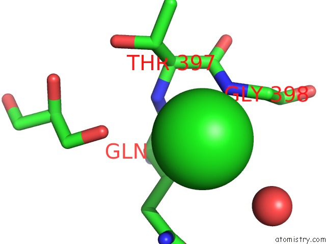
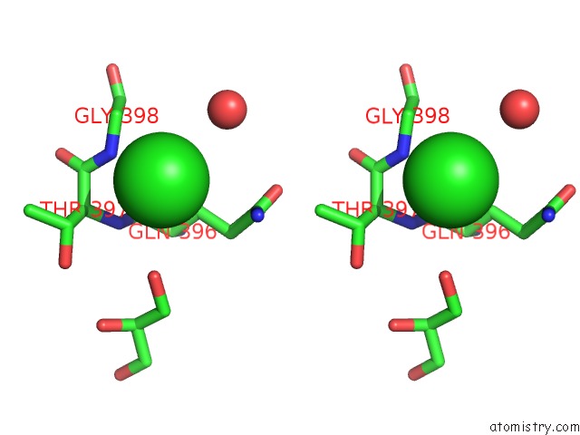
Chlorine binding site 1 out of 3 in 4xc5
Go back to
Chlorine binding site 1 out
of 3 in the Crystal Structure of the T1L Reovirus Attachment Protein SIGMA1
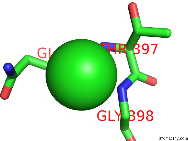
Mono view
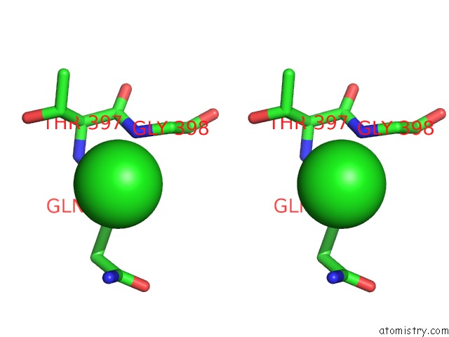
Stereo pair view
Mono view
Stereo pair view
A full contact list of Chlorine with other atoms in the Cl binding
site number 1 of Crystal Structure of the T1L Reovirus Attachment Protein SIGMA1 within 5.0Å range:
|
Chlorine binding site 2 out of 3 in 4xc5
Go back to
Chlorine binding site 2 out
of 3 in the Crystal Structure of the T1L Reovirus Attachment Protein SIGMA1
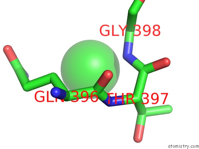
Mono view
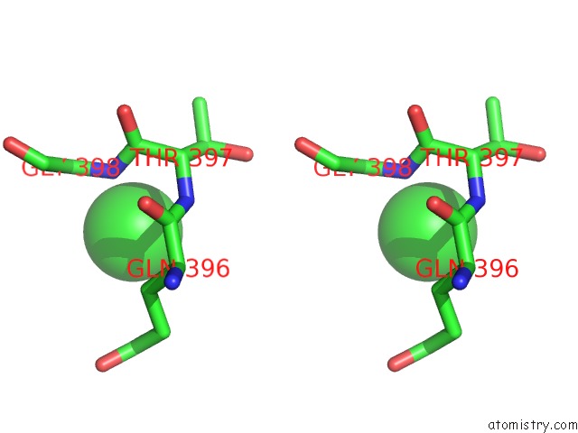
Stereo pair view
Mono view
Stereo pair view
A full contact list of Chlorine with other atoms in the Cl binding
site number 2 of Crystal Structure of the T1L Reovirus Attachment Protein SIGMA1 within 5.0Å range:
|
Chlorine binding site 3 out of 3 in 4xc5
Go back to
Chlorine binding site 3 out
of 3 in the Crystal Structure of the T1L Reovirus Attachment Protein SIGMA1
Mono view
Stereo pair view
Mono view
Stereo pair view
A full contact list of Chlorine with other atoms in the Cl binding
site number 3 of Crystal Structure of the T1L Reovirus Attachment Protein SIGMA1 within 5.0Å range:
|
Reference:
E.Stettner,
K.Reiss,
M.H.Dietrich,
K.M.Ogden,
T.S.Dermody,
T.Stehle.
Structure of Serotype 1 Reovirus Attachment Protein SIGMA1 in Complex with Jam-A Reveals A Conserved Serotype-Independent Binding Epitope To Be Published.
Page generated: Fri Jul 26 03:22:05 2024
Last articles
Zn in 9J0NZn in 9J0O
Zn in 9J0P
Zn in 9FJX
Zn in 9EKB
Zn in 9C0F
Zn in 9CAH
Zn in 9CH0
Zn in 9CH3
Zn in 9CH1