Chlorine »
PDB 4xg9-4xpb »
4xgc »
Chlorine in PDB 4xgc: Crystal Structure of the Eukaryotic Origin Recognition Complex
Enzymatic activity of Crystal Structure of the Eukaryotic Origin Recognition Complex
All present enzymatic activity of Crystal Structure of the Eukaryotic Origin Recognition Complex:
3.6.1.15;
3.6.1.15;
Protein crystallography data
The structure of Crystal Structure of the Eukaryotic Origin Recognition Complex, PDB code: 4xgc
was solved by
F.Bleichert,
M.R.Botchan,
J.M.Berger,
with X-Ray Crystallography technique. A brief refinement statistics is given in the table below:
| Resolution Low / High (Å) | 48.80 / 3.50 |
| Space group | I 2 2 2 |
| Cell size a, b, c (Å), α, β, γ (°) | 145.545, 258.983, 257.001, 90.00, 90.00, 90.00 |
| R / Rfree (%) | 22.4 / 25.8 |
Other elements in 4xgc:
The structure of Crystal Structure of the Eukaryotic Origin Recognition Complex also contains other interesting chemical elements:
| Potassium | (K) | 1 atom |
Chlorine Binding Sites:
The binding sites of Chlorine atom in the Crystal Structure of the Eukaryotic Origin Recognition Complex
(pdb code 4xgc). This binding sites where shown within
5.0 Angstroms radius around Chlorine atom.
In total only one binding site of Chlorine was determined in the Crystal Structure of the Eukaryotic Origin Recognition Complex, PDB code: 4xgc:
In total only one binding site of Chlorine was determined in the Crystal Structure of the Eukaryotic Origin Recognition Complex, PDB code: 4xgc:
Chlorine binding site 1 out of 1 in 4xgc
Go back to
Chlorine binding site 1 out
of 1 in the Crystal Structure of the Eukaryotic Origin Recognition Complex
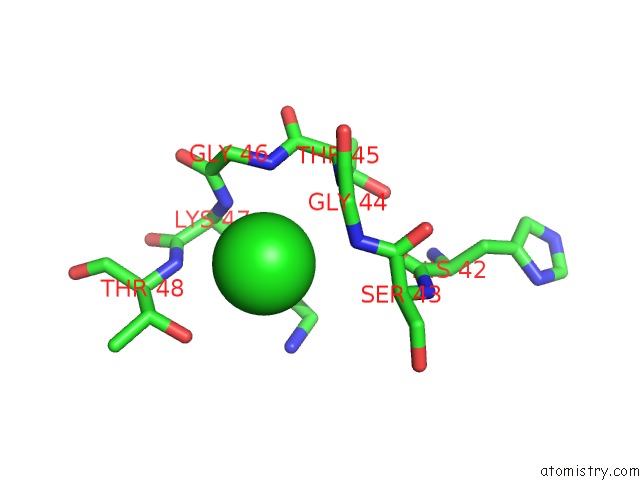
Mono view
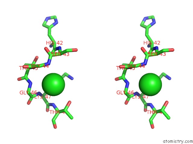
Stereo pair view
Mono view
Stereo pair view
A full contact list of Chlorine with other atoms in the Cl binding
site number 1 of Crystal Structure of the Eukaryotic Origin Recognition Complex within 5.0Å range:
|
Reference:
F.Bleichert,
M.R.Botchan,
J.M.Berger.
Crystal Structure of the Eukaryotic Origin Recognition Complex. Nature V. 519 321 2015.
ISSN: ESSN 1476-4687
PubMed: 25762138
DOI: 10.1038/NATURE14239
Page generated: Fri Jul 26 03:27:53 2024
ISSN: ESSN 1476-4687
PubMed: 25762138
DOI: 10.1038/NATURE14239
Last articles
Zn in 9J0NZn in 9J0O
Zn in 9J0P
Zn in 9FJX
Zn in 9EKB
Zn in 9C0F
Zn in 9CAH
Zn in 9CH0
Zn in 9CH3
Zn in 9CH1