Chlorine »
PDB 4y8h-4ydn »
4y9x »
Chlorine in PDB 4y9x: Crystal Structure of Glucosyl-3-Phosphoglycerate Synthase From Mycobacterium Tuberculosis in Complex with MN2+, Uridine-Diphosphate- Glucose (Udp-Glc) and Phosphoglyceric Acid (Pga) - Gpgs MN2+ Udp-Glc Pga-3
Enzymatic activity of Crystal Structure of Glucosyl-3-Phosphoglycerate Synthase From Mycobacterium Tuberculosis in Complex with MN2+, Uridine-Diphosphate- Glucose (Udp-Glc) and Phosphoglyceric Acid (Pga) - Gpgs MN2+ Udp-Glc Pga-3
All present enzymatic activity of Crystal Structure of Glucosyl-3-Phosphoglycerate Synthase From Mycobacterium Tuberculosis in Complex with MN2+, Uridine-Diphosphate- Glucose (Udp-Glc) and Phosphoglyceric Acid (Pga) - Gpgs MN2+ Udp-Glc Pga-3:
2.4.1.266;
2.4.1.266;
Protein crystallography data
The structure of Crystal Structure of Glucosyl-3-Phosphoglycerate Synthase From Mycobacterium Tuberculosis in Complex with MN2+, Uridine-Diphosphate- Glucose (Udp-Glc) and Phosphoglyceric Acid (Pga) - Gpgs MN2+ Udp-Glc Pga-3, PDB code: 4y9x
was solved by
D.Albesa-Jove,
A.Rodrigo-Unzueta,
J.O.Cifuente,
S.Urresti,
N.Comino,
E.Sancho-Vaello,
M.E.Guerin,
with X-Ray Crystallography technique. A brief refinement statistics is given in the table below:
| Resolution Low / High (Å) | 29.06 / 2.64 |
| Space group | I 41 |
| Cell size a, b, c (Å), α, β, γ (°) | 98.850, 98.850, 127.790, 90.00, 90.00, 90.00 |
| R / Rfree (%) | 21.4 / 25.2 |
Other elements in 4y9x:
The structure of Crystal Structure of Glucosyl-3-Phosphoglycerate Synthase From Mycobacterium Tuberculosis in Complex with MN2+, Uridine-Diphosphate- Glucose (Udp-Glc) and Phosphoglyceric Acid (Pga) - Gpgs MN2+ Udp-Glc Pga-3 also contains other interesting chemical elements:
| Manganese | (Mn) | 1 atom |
Chlorine Binding Sites:
The binding sites of Chlorine atom in the Crystal Structure of Glucosyl-3-Phosphoglycerate Synthase From Mycobacterium Tuberculosis in Complex with MN2+, Uridine-Diphosphate- Glucose (Udp-Glc) and Phosphoglyceric Acid (Pga) - Gpgs MN2+ Udp-Glc Pga-3
(pdb code 4y9x). This binding sites where shown within
5.0 Angstroms radius around Chlorine atom.
In total only one binding site of Chlorine was determined in the Crystal Structure of Glucosyl-3-Phosphoglycerate Synthase From Mycobacterium Tuberculosis in Complex with MN2+, Uridine-Diphosphate- Glucose (Udp-Glc) and Phosphoglyceric Acid (Pga) - Gpgs MN2+ Udp-Glc Pga-3, PDB code: 4y9x:
In total only one binding site of Chlorine was determined in the Crystal Structure of Glucosyl-3-Phosphoglycerate Synthase From Mycobacterium Tuberculosis in Complex with MN2+, Uridine-Diphosphate- Glucose (Udp-Glc) and Phosphoglyceric Acid (Pga) - Gpgs MN2+ Udp-Glc Pga-3, PDB code: 4y9x:
Chlorine binding site 1 out of 1 in 4y9x
Go back to
Chlorine binding site 1 out
of 1 in the Crystal Structure of Glucosyl-3-Phosphoglycerate Synthase From Mycobacterium Tuberculosis in Complex with MN2+, Uridine-Diphosphate- Glucose (Udp-Glc) and Phosphoglyceric Acid (Pga) - Gpgs MN2+ Udp-Glc Pga-3
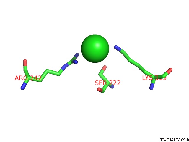
Mono view
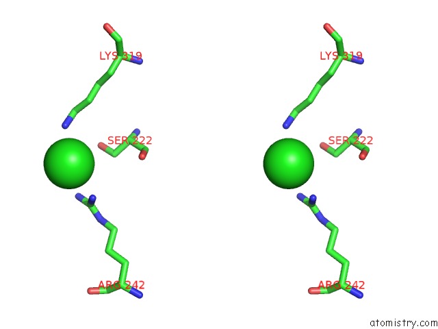
Stereo pair view
Mono view
Stereo pair view
A full contact list of Chlorine with other atoms in the Cl binding
site number 1 of Crystal Structure of Glucosyl-3-Phosphoglycerate Synthase From Mycobacterium Tuberculosis in Complex with MN2+, Uridine-Diphosphate- Glucose (Udp-Glc) and Phosphoglyceric Acid (Pga) - Gpgs MN2+ Udp-Glc Pga-3 within 5.0Å range:
|
Reference:
D.Albesa-Jove,
F.Mendoza,
A.Rodrigo-Unzueta,
F.Gomollon-Bel,
J.O.Cifuente,
S.Urresti,
N.Comino,
H.Gomez,
J.Romero-Garcia,
J.M.Lluch,
E.Sancho-Vaello,
X.Biarnes,
A.Planas,
P.Merino,
L.Masgrau,
M.E.Guerin.
A Native Ternary Complex Trapped in A Crystal Reveals the Catalytic Mechanism of A Retaining Glycosyltransferase. Angew.Chem.Int.Ed.Engl. V. 54 9898 2015.
ISSN: ESSN 1521-3773
PubMed: 26136334
DOI: 10.1002/ANIE.201504617
Page generated: Fri Jul 26 03:58:09 2024
ISSN: ESSN 1521-3773
PubMed: 26136334
DOI: 10.1002/ANIE.201504617
Last articles
Zn in 9J0NZn in 9J0O
Zn in 9J0P
Zn in 9FJX
Zn in 9EKB
Zn in 9C0F
Zn in 9CAH
Zn in 9CH0
Zn in 9CH3
Zn in 9CH1