Chlorine »
PDB 4yli-4ywy »
4ypo »
Chlorine in PDB 4ypo: Crystal Structure of Mycobacterium Tuberculosis Ketol-Acid Reductoisomerase in Complex with MG2+
Enzymatic activity of Crystal Structure of Mycobacterium Tuberculosis Ketol-Acid Reductoisomerase in Complex with MG2+
All present enzymatic activity of Crystal Structure of Mycobacterium Tuberculosis Ketol-Acid Reductoisomerase in Complex with MG2+:
1.1.1.86;
1.1.1.86;
Protein crystallography data
The structure of Crystal Structure of Mycobacterium Tuberculosis Ketol-Acid Reductoisomerase in Complex with MG2+, PDB code: 4ypo
was solved by
Y.Lv,
L.W.Guddat,
with X-Ray Crystallography technique. A brief refinement statistics is given in the table below:
| Resolution Low / High (Å) | 38.74 / 1.00 |
| Space group | C 1 2 1 |
| Cell size a, b, c (Å), α, β, γ (°) | 157.697, 54.797, 92.230, 90.00, 123.25, 90.00 |
| R / Rfree (%) | 15.5 / 16.3 |
Other elements in 4ypo:
The structure of Crystal Structure of Mycobacterium Tuberculosis Ketol-Acid Reductoisomerase in Complex with MG2+ also contains other interesting chemical elements:
| Magnesium | (Mg) | 13 atoms |
| Sodium | (Na) | 1 atom |
Chlorine Binding Sites:
The binding sites of Chlorine atom in the Crystal Structure of Mycobacterium Tuberculosis Ketol-Acid Reductoisomerase in Complex with MG2+
(pdb code 4ypo). This binding sites where shown within
5.0 Angstroms radius around Chlorine atom.
In total 2 binding sites of Chlorine where determined in the Crystal Structure of Mycobacterium Tuberculosis Ketol-Acid Reductoisomerase in Complex with MG2+, PDB code: 4ypo:
Jump to Chlorine binding site number: 1; 2;
In total 2 binding sites of Chlorine where determined in the Crystal Structure of Mycobacterium Tuberculosis Ketol-Acid Reductoisomerase in Complex with MG2+, PDB code: 4ypo:
Jump to Chlorine binding site number: 1; 2;
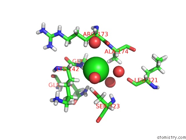
Chlorine binding site 1 out of 2 in 4ypo
Go back to
Chlorine binding site 1 out
of 2 in the Crystal Structure of Mycobacterium Tuberculosis Ketol-Acid Reductoisomerase in Complex with MG2+

Mono view

Stereo pair view
Mono view
Stereo pair view
A full contact list of Chlorine with other atoms in the Cl binding
site number 1 of Crystal Structure of Mycobacterium Tuberculosis Ketol-Acid Reductoisomerase in Complex with MG2+ within 5.0Å range:
|
Chlorine binding site 2 out of 2 in 4ypo
Go back to
Chlorine binding site 2 out
of 2 in the Crystal Structure of Mycobacterium Tuberculosis Ketol-Acid Reductoisomerase in Complex with MG2+
Mono view
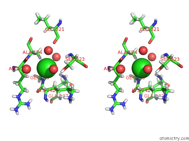
Stereo pair view
Mono view
Stereo pair view
A full contact list of Chlorine with other atoms in the Cl binding
site number 2 of Crystal Structure of Mycobacterium Tuberculosis Ketol-Acid Reductoisomerase in Complex with MG2+ within 5.0Å range:
|
Reference:
Y.Lv,
A.Kandale,
S.J.Wun,
R.P.Mcgeary,
S.J.Williams,
B.Kobe,
V.Sieber,
M.A.Schembri,
G.Schenk,
L.W.Guddat.
Crystal Structure of Mycobacterium Tuberculosis Ketol-Acid Reductoisomerase at 1.0 Angstrom Resolution - A Potential Target For Anti-Tuberculosis Drug Discovery. Febs J. V. 283 1184 2016.
ISSN: ISSN 1742-464X
PubMed: 26876563
DOI: 10.1111/FEBS.13672
Page generated: Fri Jul 26 04:13:03 2024
ISSN: ISSN 1742-464X
PubMed: 26876563
DOI: 10.1111/FEBS.13672
Last articles
Zn in 9MJ5Zn in 9HNW
Zn in 9G0L
Zn in 9FNE
Zn in 9DZN
Zn in 9E0I
Zn in 9D32
Zn in 9DAK
Zn in 8ZXC
Zn in 8ZUF