Chlorine »
PDB 4zg8-4znq »
4zli »
Chlorine in PDB 4zli: Cellobionic Acid Phosphorylase - 3-O-Beta-D-Glucopyranosyl-Alpha-D- Glucopyranuronic Acid Complex
Enzymatic activity of Cellobionic Acid Phosphorylase - 3-O-Beta-D-Glucopyranosyl-Alpha-D- Glucopyranuronic Acid Complex
All present enzymatic activity of Cellobionic Acid Phosphorylase - 3-O-Beta-D-Glucopyranosyl-Alpha-D- Glucopyranuronic Acid Complex:
2.4.1.321;
2.4.1.321;
Protein crystallography data
The structure of Cellobionic Acid Phosphorylase - 3-O-Beta-D-Glucopyranosyl-Alpha-D- Glucopyranuronic Acid Complex, PDB code: 4zli
was solved by
Y.W.Nam,
T.Arakawa,
S.Fushinobu,
with X-Ray Crystallography technique. A brief refinement statistics is given in the table below:
| Resolution Low / High (Å) | 40.50 / 1.80 |
| Space group | P 31 2 1 |
| Cell size a, b, c (Å), α, β, γ (°) | 107.058, 107.058, 185.767, 90.00, 90.00, 120.00 |
| R / Rfree (%) | 15.5 / 18.8 |
Chlorine Binding Sites:
The binding sites of Chlorine atom in the Cellobionic Acid Phosphorylase - 3-O-Beta-D-Glucopyranosyl-Alpha-D- Glucopyranuronic Acid Complex
(pdb code 4zli). This binding sites where shown within
5.0 Angstroms radius around Chlorine atom.
In total only one binding site of Chlorine was determined in the Cellobionic Acid Phosphorylase - 3-O-Beta-D-Glucopyranosyl-Alpha-D- Glucopyranuronic Acid Complex, PDB code: 4zli:
In total only one binding site of Chlorine was determined in the Cellobionic Acid Phosphorylase - 3-O-Beta-D-Glucopyranosyl-Alpha-D- Glucopyranuronic Acid Complex, PDB code: 4zli:
Chlorine binding site 1 out of 1 in 4zli
Go back to
Chlorine binding site 1 out
of 1 in the Cellobionic Acid Phosphorylase - 3-O-Beta-D-Glucopyranosyl-Alpha-D- Glucopyranuronic Acid Complex
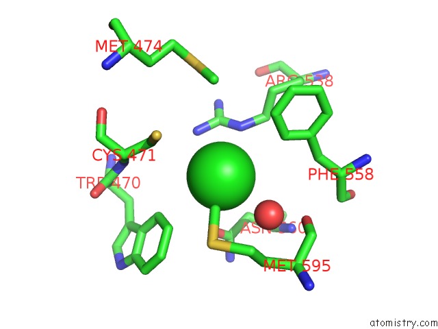
Mono view
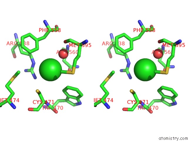
Stereo pair view
Mono view
Stereo pair view
A full contact list of Chlorine with other atoms in the Cl binding
site number 1 of Cellobionic Acid Phosphorylase - 3-O-Beta-D-Glucopyranosyl-Alpha-D- Glucopyranuronic Acid Complex within 5.0Å range:
|
Reference:
Y.W.Nam,
T.Nihira,
T.Arakawa,
Y.Saito,
M.Kitaoka,
H.Nakai,
S.Fushinobu.
Crystal Structure and Substrate Recognition of Cellobionic Acid Phosphorylase, Which Plays A Key Role in Oxidative Cellulose Degradation By Microbes. J.Biol.Chem. V. 290 18281 2015.
ISSN: ESSN 1083-351X
PubMed: 26041776
DOI: 10.1074/JBC.M115.664664
Page generated: Fri Jul 26 04:44:17 2024
ISSN: ESSN 1083-351X
PubMed: 26041776
DOI: 10.1074/JBC.M115.664664
Last articles
Zn in 9J0NZn in 9J0O
Zn in 9J0P
Zn in 9FJX
Zn in 9EKB
Zn in 9C0F
Zn in 9CAH
Zn in 9CH0
Zn in 9CH3
Zn in 9CH1