Chlorine »
PDB 4zyf-5aa2 »
5a95 »
Chlorine in PDB 5a95: Crystal Structure of Beta-Glucanase SDGLUC5_26A From Saccharophagus Degradans in Complex with Tetrasaccharide A, Form 2
Enzymatic activity of Crystal Structure of Beta-Glucanase SDGLUC5_26A From Saccharophagus Degradans in Complex with Tetrasaccharide A, Form 2
All present enzymatic activity of Crystal Structure of Beta-Glucanase SDGLUC5_26A From Saccharophagus Degradans in Complex with Tetrasaccharide A, Form 2:
3.2.1.73;
3.2.1.73;
Protein crystallography data
The structure of Crystal Structure of Beta-Glucanase SDGLUC5_26A From Saccharophagus Degradans in Complex with Tetrasaccharide A, Form 2, PDB code: 5a95
was solved by
G.Sulzenbacher,
M.Lafond,
T.Freyd,
B.Henrissat,
R.M.Coutinho,
J.G.Berrin,
M.L.Garron,
with X-Ray Crystallography technique. A brief refinement statistics is given in the table below:
| Resolution Low / High (Å) | 45.44 / 1.35 |
| Space group | P 1 21 1 |
| Cell size a, b, c (Å), α, β, γ (°) | 72.048, 60.384, 130.326, 90.00, 104.75, 90.00 |
| R / Rfree (%) | 12.9 / 15.6 |
Other elements in 5a95:
The structure of Crystal Structure of Beta-Glucanase SDGLUC5_26A From Saccharophagus Degradans in Complex with Tetrasaccharide A, Form 2 also contains other interesting chemical elements:
| Magnesium | (Mg) | 5 atoms |
| Sodium | (Na) | 1 atom |
Chlorine Binding Sites:
Pages:
>>> Page 1 <<< Page 2, Binding sites: 11 - 12;Binding sites:
The binding sites of Chlorine atom in the Crystal Structure of Beta-Glucanase SDGLUC5_26A From Saccharophagus Degradans in Complex with Tetrasaccharide A, Form 2 (pdb code 5a95). This binding sites where shown within 5.0 Angstroms radius around Chlorine atom.In total 12 binding sites of Chlorine where determined in the Crystal Structure of Beta-Glucanase SDGLUC5_26A From Saccharophagus Degradans in Complex with Tetrasaccharide A, Form 2, PDB code: 5a95:
Jump to Chlorine binding site number: 1; 2; 3; 4; 5; 6; 7; 8; 9; 10;
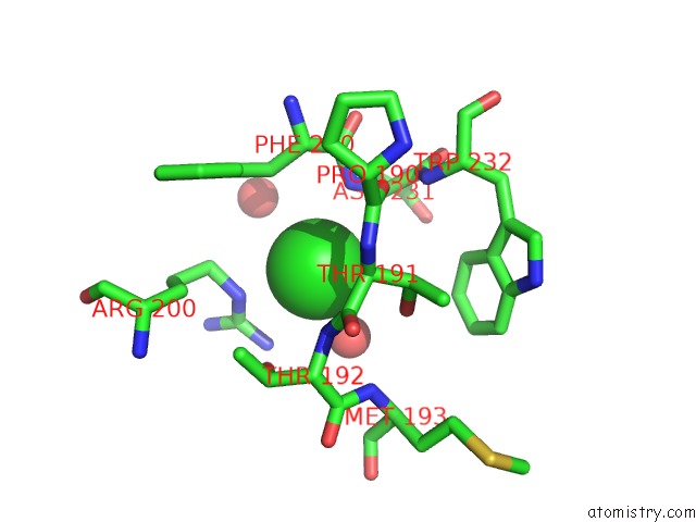
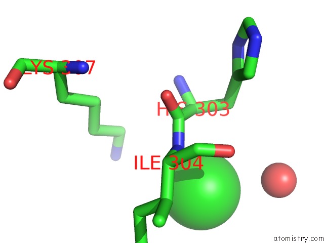
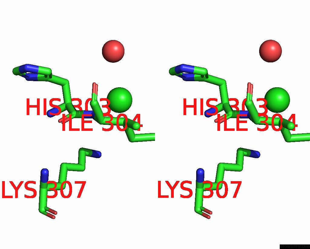
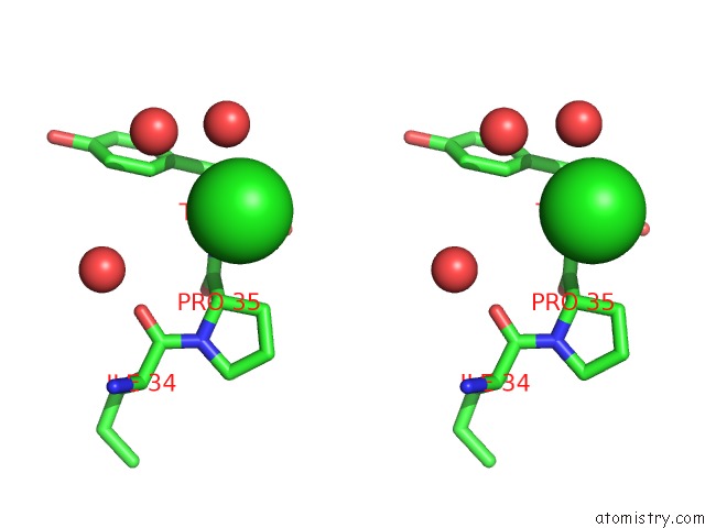
Chlorine binding site 1 out of 12 in 5a95
Go back to
Chlorine binding site 1 out
of 12 in the Crystal Structure of Beta-Glucanase SDGLUC5_26A From Saccharophagus Degradans in Complex with Tetrasaccharide A, Form 2
Mono view

Stereo pair view
Mono view
Stereo pair view
A full contact list of Chlorine with other atoms in the Cl binding
site number 1 of Crystal Structure of Beta-Glucanase SDGLUC5_26A From Saccharophagus Degradans in Complex with Tetrasaccharide A, Form 2 within 5.0Å range:
|
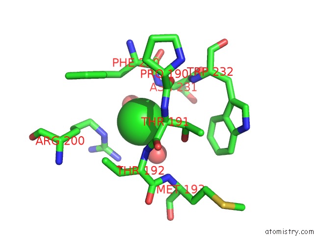
Chlorine binding site 2 out of 12 in 5a95
Go back to
Chlorine binding site 2 out
of 12 in the Crystal Structure of Beta-Glucanase SDGLUC5_26A From Saccharophagus Degradans in Complex with Tetrasaccharide A, Form 2
Mono view

Stereo pair view
Mono view
Stereo pair view
A full contact list of Chlorine with other atoms in the Cl binding
site number 2 of Crystal Structure of Beta-Glucanase SDGLUC5_26A From Saccharophagus Degradans in Complex with Tetrasaccharide A, Form 2 within 5.0Å range:
|
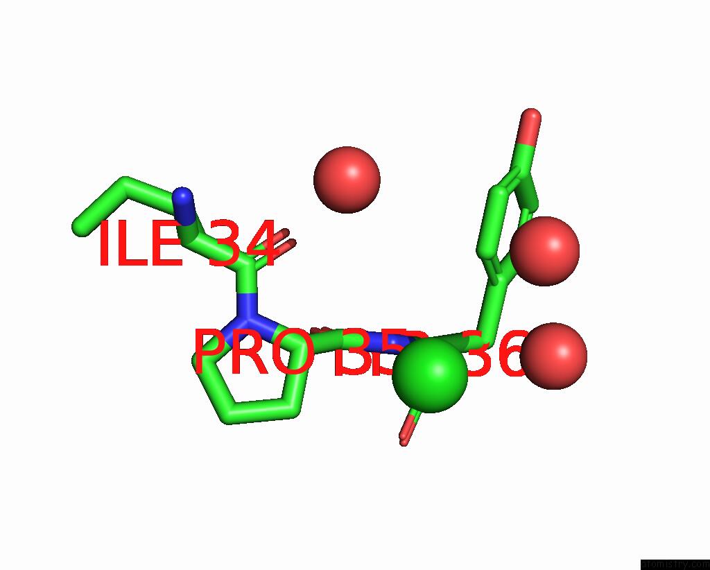
Chlorine binding site 3 out of 12 in 5a95
Go back to
Chlorine binding site 3 out
of 12 in the Crystal Structure of Beta-Glucanase SDGLUC5_26A From Saccharophagus Degradans in Complex with Tetrasaccharide A, Form 2
Mono view
Stereo pair view
Mono view
Stereo pair view
A full contact list of Chlorine with other atoms in the Cl binding
site number 3 of Crystal Structure of Beta-Glucanase SDGLUC5_26A From Saccharophagus Degradans in Complex with Tetrasaccharide A, Form 2 within 5.0Å range:
|
Chlorine binding site 4 out of 12 in 5a95
Go back to
Chlorine binding site 4 out
of 12 in the Crystal Structure of Beta-Glucanase SDGLUC5_26A From Saccharophagus Degradans in Complex with Tetrasaccharide A, Form 2
Mono view
Stereo pair view
Mono view
Stereo pair view
A full contact list of Chlorine with other atoms in the Cl binding
site number 4 of Crystal Structure of Beta-Glucanase SDGLUC5_26A From Saccharophagus Degradans in Complex with Tetrasaccharide A, Form 2 within 5.0Å range:
|
Chlorine binding site 5 out of 12 in 5a95
Go back to
Chlorine binding site 5 out
of 12 in the Crystal Structure of Beta-Glucanase SDGLUC5_26A From Saccharophagus Degradans in Complex with Tetrasaccharide A, Form 2
Mono view

Stereo pair view
Mono view
Stereo pair view
A full contact list of Chlorine with other atoms in the Cl binding
site number 5 of Crystal Structure of Beta-Glucanase SDGLUC5_26A From Saccharophagus Degradans in Complex with Tetrasaccharide A, Form 2 within 5.0Å range:
|
Chlorine binding site 6 out of 12 in 5a95
Go back to
Chlorine binding site 6 out
of 12 in the Crystal Structure of Beta-Glucanase SDGLUC5_26A From Saccharophagus Degradans in Complex with Tetrasaccharide A, Form 2

Mono view

Stereo pair view
Mono view
Stereo pair view
A full contact list of Chlorine with other atoms in the Cl binding
site number 6 of Crystal Structure of Beta-Glucanase SDGLUC5_26A From Saccharophagus Degradans in Complex with Tetrasaccharide A, Form 2 within 5.0Å range:
|
Chlorine binding site 7 out of 12 in 5a95
Go back to
Chlorine binding site 7 out
of 12 in the Crystal Structure of Beta-Glucanase SDGLUC5_26A From Saccharophagus Degradans in Complex with Tetrasaccharide A, Form 2
Mono view
Stereo pair view
Mono view
Stereo pair view
A full contact list of Chlorine with other atoms in the Cl binding
site number 7 of Crystal Structure of Beta-Glucanase SDGLUC5_26A From Saccharophagus Degradans in Complex with Tetrasaccharide A, Form 2 within 5.0Å range:
|
Chlorine binding site 8 out of 12 in 5a95
Go back to
Chlorine binding site 8 out
of 12 in the Crystal Structure of Beta-Glucanase SDGLUC5_26A From Saccharophagus Degradans in Complex with Tetrasaccharide A, Form 2
Mono view

Stereo pair view
Mono view
Stereo pair view
A full contact list of Chlorine with other atoms in the Cl binding
site number 8 of Crystal Structure of Beta-Glucanase SDGLUC5_26A From Saccharophagus Degradans in Complex with Tetrasaccharide A, Form 2 within 5.0Å range:
|
Chlorine binding site 9 out of 12 in 5a95
Go back to
Chlorine binding site 9 out
of 12 in the Crystal Structure of Beta-Glucanase SDGLUC5_26A From Saccharophagus Degradans in Complex with Tetrasaccharide A, Form 2
Mono view

Stereo pair view
Mono view
Stereo pair view
A full contact list of Chlorine with other atoms in the Cl binding
site number 9 of Crystal Structure of Beta-Glucanase SDGLUC5_26A From Saccharophagus Degradans in Complex with Tetrasaccharide A, Form 2 within 5.0Å range:
|
Chlorine binding site 10 out of 12 in 5a95
Go back to
Chlorine binding site 10 out
of 12 in the Crystal Structure of Beta-Glucanase SDGLUC5_26A From Saccharophagus Degradans in Complex with Tetrasaccharide A, Form 2

Mono view

Stereo pair view
Mono view
Stereo pair view
A full contact list of Chlorine with other atoms in the Cl binding
site number 10 of Crystal Structure of Beta-Glucanase SDGLUC5_26A From Saccharophagus Degradans in Complex with Tetrasaccharide A, Form 2 within 5.0Å range:
|
Reference:
M.Lafond,
G.Sulzenbacher,
T.Freyd,
B.Henrissat,
J.G.Berrin,
M.L.Garron.
The Quaternary Structure of A Glycoside Hydrolase Dictates Specificity Towards Beta-Glucans J.Biol.Chem. V. 291 7183 2016.
ISSN: ISSN 0021-9258
PubMed: 26755730
DOI: 10.1074/JBC.M115.695999
Page generated: Fri Jul 26 05:01:52 2024
ISSN: ISSN 0021-9258
PubMed: 26755730
DOI: 10.1074/JBC.M115.695999
Last articles
Zn in 9J0NZn in 9J0O
Zn in 9J0P
Zn in 9FJX
Zn in 9EKB
Zn in 9C0F
Zn in 9CAH
Zn in 9CH0
Zn in 9CH3
Zn in 9CH1