Chlorine »
PDB 5afy-5anv »
5ale »
Chlorine in PDB 5ale: Ligand Complex Structure of Soluble Epoxide Hydrolase
Enzymatic activity of Ligand Complex Structure of Soluble Epoxide Hydrolase
All present enzymatic activity of Ligand Complex Structure of Soluble Epoxide Hydrolase:
3.1.3.76; 3.3.2.10;
3.1.3.76; 3.3.2.10;
Protein crystallography data
The structure of Ligand Complex Structure of Soluble Epoxide Hydrolase, PDB code: 5ale
was solved by
L.Oster,
S.Tapani,
Y.Xue,
H.Kack,
with X-Ray Crystallography technique. A brief refinement statistics is given in the table below:
| Resolution Low / High (Å) | 48.48 / 1.95 |
| Space group | P 65 2 2 |
| Cell size a, b, c (Å), α, β, γ (°) | 92.320, 92.320, 243.906, 90.00, 90.00, 120.00 |
| R / Rfree (%) | 19.6 / 23.08 |
Chlorine Binding Sites:
The binding sites of Chlorine atom in the Ligand Complex Structure of Soluble Epoxide Hydrolase
(pdb code 5ale). This binding sites where shown within
5.0 Angstroms radius around Chlorine atom.
In total only one binding site of Chlorine was determined in the Ligand Complex Structure of Soluble Epoxide Hydrolase, PDB code: 5ale:
In total only one binding site of Chlorine was determined in the Ligand Complex Structure of Soluble Epoxide Hydrolase, PDB code: 5ale:
Chlorine binding site 1 out of 1 in 5ale
Go back to
Chlorine binding site 1 out
of 1 in the Ligand Complex Structure of Soluble Epoxide Hydrolase
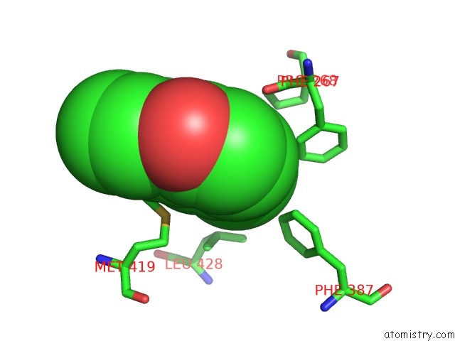
Mono view
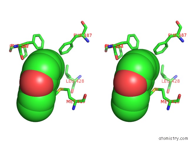
Stereo pair view
Mono view
Stereo pair view
A full contact list of Chlorine with other atoms in the Cl binding
site number 1 of Ligand Complex Structure of Soluble Epoxide Hydrolase within 5.0Å range:
|
Reference:
L.Oster,
S.Tapani,
Y.Xue,
H.Kack.
Successful Generation of Structural Information For Fragment-Based Drug Discovery. Drug Discov Today 2015.
ISSN: ESSN 1878-5832
PubMed: 25931264
DOI: 10.1016/J.DRUDIS.2015.04.005
Page generated: Fri Jul 26 05:18:23 2024
ISSN: ESSN 1878-5832
PubMed: 25931264
DOI: 10.1016/J.DRUDIS.2015.04.005
Last articles
Zn in 9JYWZn in 9IR4
Zn in 9IR3
Zn in 9GMX
Zn in 9GMW
Zn in 9JEJ
Zn in 9ERF
Zn in 9ERE
Zn in 9EGV
Zn in 9EGW