Chlorine »
PDB 5aoa-5b1k »
5b07 »
Chlorine in PDB 5b07: Lysozyme (Denatured By Dcl and Refolded)
Enzymatic activity of Lysozyme (Denatured By Dcl and Refolded)
All present enzymatic activity of Lysozyme (Denatured By Dcl and Refolded):
3.2.1.17;
3.2.1.17;
Protein crystallography data
The structure of Lysozyme (Denatured By Dcl and Refolded), PDB code: 5b07
was solved by
A.Kita,
Y.Morimoto,
with X-Ray Crystallography technique. A brief refinement statistics is given in the table below:
| Resolution Low / High (Å) | 55.73 / 1.80 |
| Space group | P 43 21 2 |
| Cell size a, b, c (Å), α, β, γ (°) | 78.816, 78.816, 36.800, 90.00, 90.00, 90.00 |
| R / Rfree (%) | 15 / 18.7 |
Other elements in 5b07:
The structure of Lysozyme (Denatured By Dcl and Refolded) also contains other interesting chemical elements:
| Sodium | (Na) | 8 atoms |
Chlorine Binding Sites:
The binding sites of Chlorine atom in the Lysozyme (Denatured By Dcl and Refolded)
(pdb code 5b07). This binding sites where shown within
5.0 Angstroms radius around Chlorine atom.
In total 4 binding sites of Chlorine where determined in the Lysozyme (Denatured By Dcl and Refolded), PDB code: 5b07:
Jump to Chlorine binding site number: 1; 2; 3; 4;
In total 4 binding sites of Chlorine where determined in the Lysozyme (Denatured By Dcl and Refolded), PDB code: 5b07:
Jump to Chlorine binding site number: 1; 2; 3; 4;
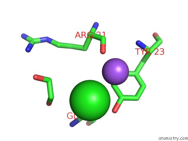
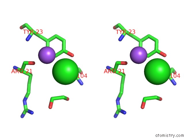
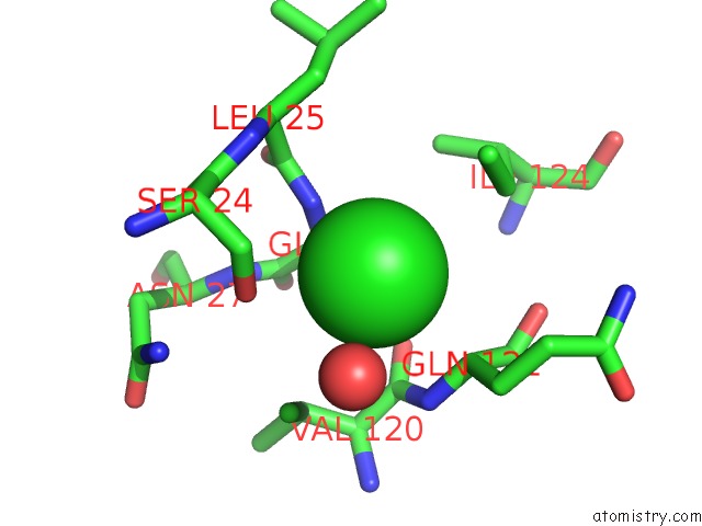
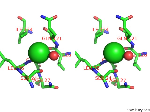
Chlorine binding site 1 out of 4 in 5b07
Go back to
Chlorine binding site 1 out
of 4 in the Lysozyme (Denatured By Dcl and Refolded)
Mono view
Stereo pair view
Mono view
Stereo pair view
A full contact list of Chlorine with other atoms in the Cl binding
site number 1 of Lysozyme (Denatured By Dcl and Refolded) within 5.0Å range:
|
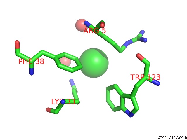
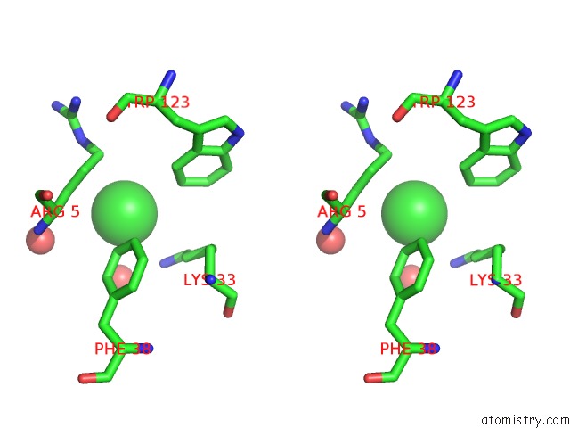
Chlorine binding site 2 out of 4 in 5b07
Go back to
Chlorine binding site 2 out
of 4 in the Lysozyme (Denatured By Dcl and Refolded)
Mono view
Stereo pair view
Mono view
Stereo pair view
A full contact list of Chlorine with other atoms in the Cl binding
site number 2 of Lysozyme (Denatured By Dcl and Refolded) within 5.0Å range:
|
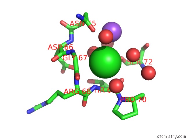
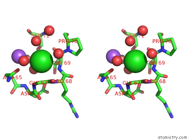
Chlorine binding site 3 out of 4 in 5b07
Go back to
Chlorine binding site 3 out
of 4 in the Lysozyme (Denatured By Dcl and Refolded)
Mono view
Stereo pair view
Mono view
Stereo pair view
A full contact list of Chlorine with other atoms in the Cl binding
site number 3 of Lysozyme (Denatured By Dcl and Refolded) within 5.0Å range:
|
Chlorine binding site 4 out of 4 in 5b07
Go back to
Chlorine binding site 4 out
of 4 in the Lysozyme (Denatured By Dcl and Refolded)
Mono view
Stereo pair view
Mono view
Stereo pair view
A full contact list of Chlorine with other atoms in the Cl binding
site number 4 of Lysozyme (Denatured By Dcl and Refolded) within 5.0Å range:
|
Reference:
A.Kita,
Y.Morimoto.
An Effective Deuterium Exchange Method For Neutron Crystal Structure Analysis with Unfolding-Refolding Processes Mol Biotechnol. V. 58 130 2016.
ISSN: ESSN 1559-0305
PubMed: 26718545
DOI: 10.1007/S12033-015-9908-8
Page generated: Fri Jul 26 05:26:42 2024
ISSN: ESSN 1559-0305
PubMed: 26718545
DOI: 10.1007/S12033-015-9908-8
Last articles
Zn in 9J0NZn in 9J0O
Zn in 9J0P
Zn in 9FJX
Zn in 9EKB
Zn in 9C0F
Zn in 9CAH
Zn in 9CH0
Zn in 9CH3
Zn in 9CH1