Chlorine »
PDB 5ao9-5b1g »
5b18 »
Chlorine in PDB 5b18: Crystal Structure of A Darunavir Resistant Hiv-1 Protease
Protein crystallography data
The structure of Crystal Structure of A Darunavir Resistant Hiv-1 Protease, PDB code: 5b18
was solved by
K.Suzuki,
H.Ode,
M.Nakashima,
W.Sugiura,
N.Watanabe,
A.Suzuki,
Y.Iwatani,
with X-Ray Crystallography technique. A brief refinement statistics is given in the table below:
| Resolution Low / High (Å) | 50.00 / 1.80 |
| Space group | P 1 |
| Cell size a, b, c (Å), α, β, γ (°) | 37.149, 48.699, 54.151, 89.90, 73.98, 86.69 |
| R / Rfree (%) | 19.3 / 24.4 |
Chlorine Binding Sites:
The binding sites of Chlorine atom in the Crystal Structure of A Darunavir Resistant Hiv-1 Protease
(pdb code 5b18). This binding sites where shown within
5.0 Angstroms radius around Chlorine atom.
In total 4 binding sites of Chlorine where determined in the Crystal Structure of A Darunavir Resistant Hiv-1 Protease, PDB code: 5b18:
Jump to Chlorine binding site number: 1; 2; 3; 4;
In total 4 binding sites of Chlorine where determined in the Crystal Structure of A Darunavir Resistant Hiv-1 Protease, PDB code: 5b18:
Jump to Chlorine binding site number: 1; 2; 3; 4;
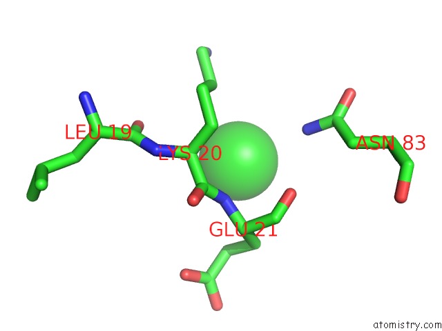
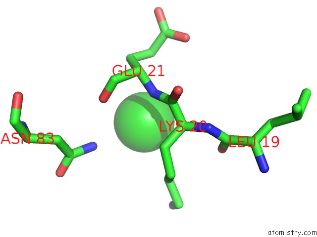
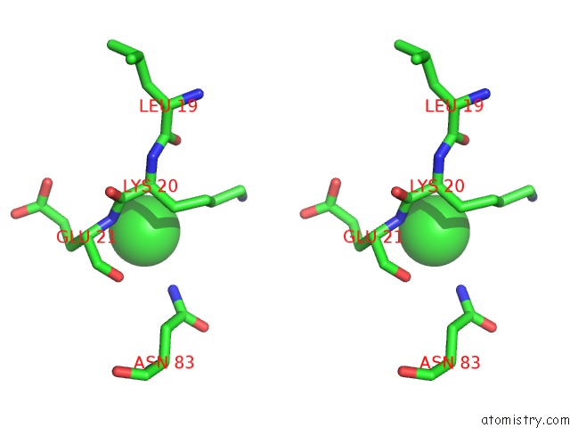
Chlorine binding site 1 out of 4 in 5b18
Go back to
Chlorine binding site 1 out
of 4 in the Crystal Structure of A Darunavir Resistant Hiv-1 Protease
Mono view
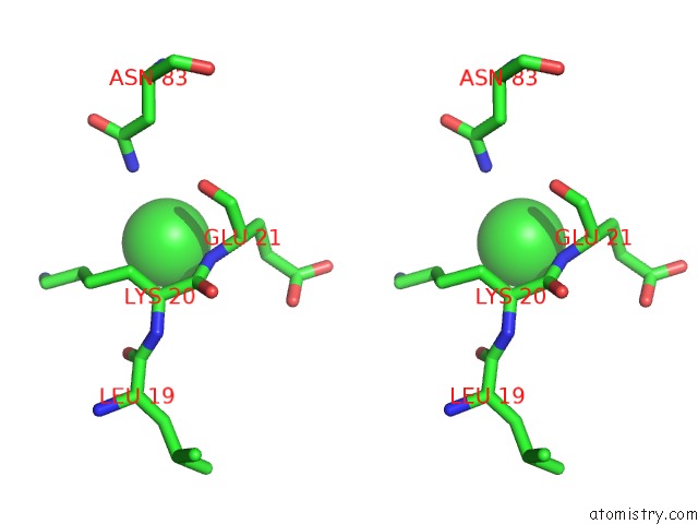
Stereo pair view
Mono view
Stereo pair view
A full contact list of Chlorine with other atoms in the Cl binding
site number 1 of Crystal Structure of A Darunavir Resistant Hiv-1 Protease within 5.0Å range:
|
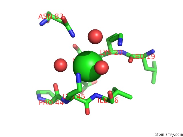
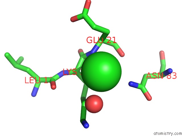
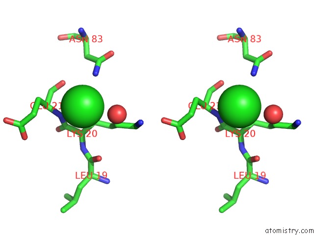
Chlorine binding site 2 out of 4 in 5b18
Go back to
Chlorine binding site 2 out
of 4 in the Crystal Structure of A Darunavir Resistant Hiv-1 Protease
Mono view
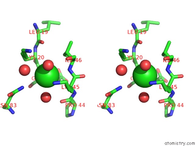
Stereo pair view
Mono view
Stereo pair view
A full contact list of Chlorine with other atoms in the Cl binding
site number 2 of Crystal Structure of A Darunavir Resistant Hiv-1 Protease within 5.0Å range:
|
Chlorine binding site 3 out of 4 in 5b18
Go back to
Chlorine binding site 3 out
of 4 in the Crystal Structure of A Darunavir Resistant Hiv-1 Protease
Mono view
Stereo pair view
Mono view
Stereo pair view
A full contact list of Chlorine with other atoms in the Cl binding
site number 3 of Crystal Structure of A Darunavir Resistant Hiv-1 Protease within 5.0Å range:
|
Chlorine binding site 4 out of 4 in 5b18
Go back to
Chlorine binding site 4 out
of 4 in the Crystal Structure of A Darunavir Resistant Hiv-1 Protease
Mono view
Stereo pair view
Mono view
Stereo pair view
A full contact list of Chlorine with other atoms in the Cl binding
site number 4 of Crystal Structure of A Darunavir Resistant Hiv-1 Protease within 5.0Å range:
|
Reference:
M.Nakashima,
H.Ode,
K.Suzuki,
M.Fujino,
M.Maejima,
Y.Kimura,
T.Masaoka,
J.Hattori,
M.Matsuda,
A.Hachiya,
Y.Yokomaku,
A.Suzuki,
N.Watanabe,
W.Sugiura,
Y.Iwatani.
Unique Flap Conformation in An Hiv-1 Protease with High-Level Darunavir Resistance Front Microbiol V. 7 61 2016.
ISSN: ESSN 1664-302X
PubMed: 26870021
DOI: 10.3389/FMICB.2016.00061
Page generated: Fri Jul 26 05:27:33 2024
ISSN: ESSN 1664-302X
PubMed: 26870021
DOI: 10.3389/FMICB.2016.00061
Last articles
Zn in 9JYWZn in 9IR4
Zn in 9IR3
Zn in 9GMX
Zn in 9GMW
Zn in 9JEJ
Zn in 9ERF
Zn in 9ERE
Zn in 9EGV
Zn in 9EGW