Chlorine »
PDB 5b1l-5bou »
5b60 »
Chlorine in PDB 5b60: Crystal Structure of PTLCIB4 S47R Mutant, A Homolog of the Limiting CO2-Inducible Protein Lcib
Protein crystallography data
The structure of Crystal Structure of PTLCIB4 S47R Mutant, A Homolog of the Limiting CO2-Inducible Protein Lcib, PDB code: 5b60
was solved by
S.Jin,
J.Sun,
T.Wunder,
D.Tang,
O.M.Mueller-Caja,
Y.Gao,
with X-Ray Crystallography technique. A brief refinement statistics is given in the table below:
| Resolution Low / High (Å) | 47.16 / 2.20 |
| Space group | C 1 2 1 |
| Cell size a, b, c (Å), α, β, γ (°) | 68.550, 66.110, 69.060, 90.00, 101.00, 90.00 |
| R / Rfree (%) | 25 / 25.4 |
Other elements in 5b60:
The structure of Crystal Structure of PTLCIB4 S47R Mutant, A Homolog of the Limiting CO2-Inducible Protein Lcib also contains other interesting chemical elements:
| Zinc | (Zn) | 1 atom |
Chlorine Binding Sites:
The binding sites of Chlorine atom in the Crystal Structure of PTLCIB4 S47R Mutant, A Homolog of the Limiting CO2-Inducible Protein Lcib
(pdb code 5b60). This binding sites where shown within
5.0 Angstroms radius around Chlorine atom.
In total only one binding site of Chlorine was determined in the Crystal Structure of PTLCIB4 S47R Mutant, A Homolog of the Limiting CO2-Inducible Protein Lcib, PDB code: 5b60:
In total only one binding site of Chlorine was determined in the Crystal Structure of PTLCIB4 S47R Mutant, A Homolog of the Limiting CO2-Inducible Protein Lcib, PDB code: 5b60:
Chlorine binding site 1 out of 1 in 5b60
Go back to
Chlorine binding site 1 out
of 1 in the Crystal Structure of PTLCIB4 S47R Mutant, A Homolog of the Limiting CO2-Inducible Protein Lcib
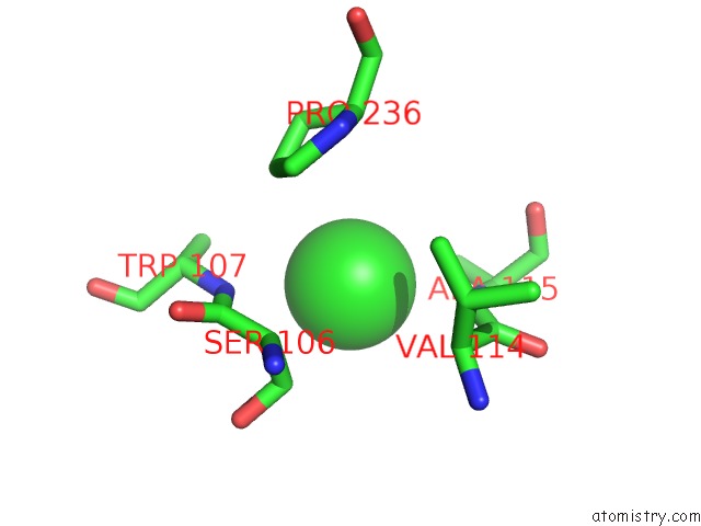
Mono view
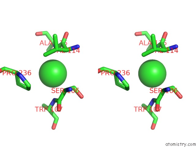
Stereo pair view
Mono view
Stereo pair view
A full contact list of Chlorine with other atoms in the Cl binding
site number 1 of Crystal Structure of PTLCIB4 S47R Mutant, A Homolog of the Limiting CO2-Inducible Protein Lcib within 5.0Å range:
|
Reference:
S.Jin,
J.Sun,
T.Wunder,
D.Tang,
A.B.Cousins,
S.K.Sze,
O.Mueller-Cajar,
Y.G.Gao.
Structural Insights Into the Lcib Protein Family Reveals A New Group of Beta-Carbonic Anhydrases Proc. Natl. Acad. Sci. V. 113 14716 2016U.S.A..
ISSN: ESSN 1091-6490
PubMed: 27911826
DOI: 10.1073/PNAS.1616294113
Page generated: Fri Jul 26 05:32:24 2024
ISSN: ESSN 1091-6490
PubMed: 27911826
DOI: 10.1073/PNAS.1616294113
Last articles
Zn in 9J0NZn in 9J0O
Zn in 9J0P
Zn in 9FJX
Zn in 9EKB
Zn in 9C0F
Zn in 9CAH
Zn in 9CH0
Zn in 9CH3
Zn in 9CH1